Evaluation of Urine May Aid in the Diagnosis of Lambda Light Chain Myeloma

SUMMARY: Multiple Myeloma is a clonal disorder of plasma cells in the bone marrow and the American Cancer Society estimates that in the United States, 30,770 new cases will be diagnosed in 2018 and 12,770 patients are expected to die of the disease. Multiple Myeloma (MM) in 2018 remains an incurable disease. The therapeutic goal therefore is to improve Progression Free Survival (PFS) and Overall Survival (OS). Multiple Myeloma evolves from a precursor stage called Monoclonal Gammopathy of Undetermined Significance (MGUS) to MM. Smoldering Multiple Myeloma (SMM) is an intermediate stage in this process of disease evolution.

Monoclonal gammopathies are often diagnosed by electrophoretic examination of the serum and urine proteins. They include Serum Protein ElectroPhoresis (SPEP), Serum Protein Immunofixation electrophoresis (SIFE), Urine Protein ElectroPhoresis (UPEP), Urine Protein Immunofixation electrophoresis (UIFE), and Bone Marrow evaluation. In addition, quantitative assessment of Serum Free Light Chains has been recommended for diagnosis and monitoring of neoplastic monoclonal gammopathies. Normal-Immunoglobulin

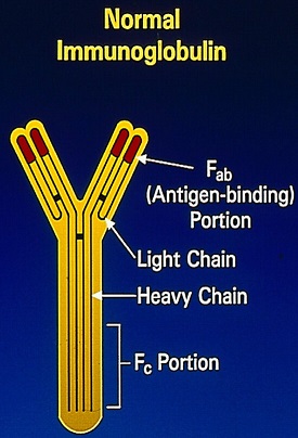
The Y-shaped Immunoglobulins are heterodimeric proteins composed of two heavy (H) and two light (L) chains. The two different types of light chains include Kappa and Lambda, which are distinctive in their amino acid sequence, with normally twice as many Kappa light chains. Malignancy can affect both types of light chains and in Multiple Myeloma. The relevant light chain production can increase, but the increase is more often in the Kappa light chains. Immunoglobulin light chains are produced in excess of the corresponding heavy chains and the excess free light chains can be quantified in serum and can also be detected in the urine, as they, by virtue of their size, are freely filtered through the glomerulus (Bence Jones protein). Excess amounts of free monoclonal light chains in patients with monoclonal gammopathy can produce nephropathy due to precipitation of these proteins in renal tubules.

Serum free Kappa and Lambda light chains are normally present in a ratio of about 0.26 to 1.65 and this ratio is increased in patients with Kappa light chain monoclonal gammopathy and decreased in patients with Lambda chain producing monoclonal gammopathies. Even though alteration in the serum K/L ratio is an important diagnostic criterion for plasma cell neoplasms, there is a high rate of positive results in patients receiving tertiary care, with abnormal K/L ratio noted in 36% of patients and about 90% of these are Kappa light chain dominant.

The serum K/L ratio is however less frequently abnormal and stays normal in patients with Lambda chain lesions even when an abnormal Lambda immunoglobulin is detected in the urine. These variabilities can result in the less-common Lambda chain-associated lesions going undiagnosed. There is a high false negative rate for Lambda dominant K/L ratio in Lambda chain associated monoclonal gammopathy (89% for MGUS, 60% for SMM and 51% for MM). It is estimated that the overall excess false negative K/L ratio rate for Lambda chain lesions, compared to Kappa chain lesions, is approximately 30%. The high false negative rate for the Lambda dominant K/L ratio, in patients with Lambda chain neoplastic monoclonal gammopathies, may be due to under-detection of Lambda light chains, Lambda chains are not produced in as much excess as are Kappa chains resulting in lower rates of Lambda dominant K/L ratio in patient with Lambda light chain neoplastic monoclonal gammopathy, and overproduction of polyclonal Kappa light chains in Lambda chain monoclonal gammopathies, as is usually noted in patients receiving tertiary care.

This study was undertaken by comparing the results of Serum and Urine Protein Electrophoreses with the results of Serum Free Light Chain Assay (SFLCA), to ascertain if the levels of overproduction of the Kappa and Lambda light chain types and their detection rates are different in patients with neoplastic monoclonal gammopathies. The authors performed a retrospective review of SPEP/SIFE, UPEP/UIFE, and SFLCA results from January 2010 through September 2017 from a total of 482 patients. Among these patients, 175 patients had a diagnosis of Neoplastic Monoclonal Gammopathy (MGUS, SMM or MM). , and evaluable results were available to address the questions of this study. Patients with Lymphomas and CLL were excluded.

The authors noted that the serum K/L ratios were appropriately abnormal more often in Kappa light chain disease. In contrast, in those with Lambda light chain disease, the K/L ratios were normal in about 25% of patients but free homogenous Lambda light chains were detectable in urine.

It was concluded that the serum Kappa/Lambda ratio in patients with Lambda light chain disease can be normal in a 25% of patients with Neoplastic Monoclonal Gammopathy and can be missed if not further evaluated with UPEP/UIFE. The authors comment that UPEP/UIFE is under- utilized and the study results question the medical necessity and clinical usefulness of the serum free light chain assay. Serum Free Light Chains in Neoplastic Monoclonal Gammopathies: Relative Under-Detection of Lambda Dominant Kappa/Lambda Ratio, and Underproduction of Free Lambda Light Chains, as Compared to Kappa Light Chains, in Patients With Neoplastic Monoclonal Gammopathies. Lee WS and Singh G. J Clin Med Res. 2018;10:562-569.

First Line KEYTRUDA® plus Chemotherapy Improves Overall Survival in Advanced Squamous NSCLC

SUMMARY: Lung cancer is the second most common cancer in both men and women and accounts for about 14% of all new cancers and 27% of all cancer deaths. The American Cancer Society estimates that for 2018 about 234,030 new cases of lung cancer will be diagnosed and over 154,050 patients will die of the disease. Lung cancer is the leading cause of cancer-related mortality in the United States. Non Small Cell Lung Cancer (NSCLC) accounts for approximately 85% of all lung cancers. Of the three main subtypes of Non Small Cell Lung Cancer (NSCLC), approximately 30% are Squamous Cell Carcinomas (SCC), 40% are Adenocarcinomas, and 10% are Large cell carcinomas. Non Small Cell Lung Cancer patients with Squamous Cell histology have been a traditionally hard- to-treat, patient group, and less than 15% of patients with advanced Squamous NSCLC survive a year after diagnosis and less than 5% of patients survive for five years or longer.

KEYTRUDA® (Pembrolizumab) is a fully humanized, Immunoglobulin G4, anti-PD-1, monoclonal antibody, that binds to the PD-1 receptor and blocks its interaction with ligands PD-L1 and PD-L2. It thereby reverses the PD-1 pathway-mediated inhibition of the immune response and unleashes the tumor-specific effector T cells. The FDA approved KEYTRUDA® for the first-line treatment of advanced NSCLC with high PD-L1 expression (Tumor Proportion Score of 50% or more), as well as in combination with Pemetrexed and Carboplatin, as first-line treatment of patients with metastatic nonsquamous NSCLC and for previously treated advanced NSCLC with a PD-L1 Tumor Proportion Score of 1% or more. Currently, KEYTRUDA® currently is the only FDA approved immunotherapy for initial treatment of NSCLC as monotherapy (KEYNOTE-024) or in combination with chemotherapy (KEYNOTE-189).Unleashing-T-Cell-Function-with-Combination-Immunotherapy

KEYNOTE-407 is a global, double-blind, placebo-controlled, phase 3 trial which compared KEYTRUDA® plus chemotherapy with placebo plus chemotherapy in patients with squamous NSCLC of any level of PD-L1 expression. In this study, 559 patients with untreated metastatic, squamous NSCLC were randomly assigned, in a 1:1 ratio to receive KEYTRUDA® 200 mg IV along with Carboplatin AUC 6 and either TAXOL® (Paclitaxel) 200 mg/m2 IV or ABRAXANE® (nab-paclitaxel) 100 mg/m2 IV days 1, 8 and 15. every 3 weeks for 4 cycles (N=278) or placebo with the same chemotherapy regimen for 4 cycles (N=281). Patients in the experimental arm following the first 4 cycles continued KEYTRUDA® every 3 weeks, for an additional 31 cycles, whereas the control group received placebo. Patients in the placebo-combination group were eligible to cross over to receive KEYTRUDA® monotherapy and 42.8% of patients assigned to the placebo plus chemotherapy group who discontinued chemotherapy went on to receive subsequent anti PD-1 or anti PD-L1 therapy and 75 patients received KEYTRUDA® monotherapy as part of in-study crossover. Patients were stratified according to the PD-L1 Tumor Proportion Score (1% or less versus more than 1%), choice of Taxane (Paclitaxel versus nab-Paclitaxel), and geographic region of enrollment. Tumor Proportion Score is the percentage of tumor cells with membranous PD-L1 staining, with less than 1% indicating PD-L1 negative score. Both treatment groups were well balanced. The co-Primary end points were Overall Survival and Progression Free Survival and the Secondary end points included Response Rate, Duration of Response and Safety. The effects of PD-L1 expression on Overall Survival (OS), Progression Free Survival (PFS), and Objective Response Rate (ORR) were prespecified exploratory end points.

At the second interim analysis, after a median follow-up of 7.8 months, the median Overall Survival was 15.9 months in the KEYTRUDA® combination group and 11.3 months in the placebo combination group (HR=0.64; P<0.001). This meant that there was a 36% reduction in the risk of death with the addition of KEYTRUDA® to chemotherapy. This OS benefit was consistently seen regardless of the level of PD-L1 expression. The median PFS was 6.4 months in the KEYTRUDA® combination group and 4.8 months in the placebo combination group (HR=0.56; P<0.001) and this suggested that the risk of disease progression or death was 44% lower with the addition of KEYTRUDA® to chemotherapy. The PFS benefit with the addition of KEYTRUDA® to chemotherapy was observed in all prespecified subgroups with incremental improvements noted with increasing PD-L1 Tumor Proportion Score. The Objective Response Rate was also significantly higher in the KEYTRUDA® chemotherapy group compared to the placebo chemotherapy group (59.4% versus 38%; P=0.0004), with a median time to response of 1.4 months and median Duration of Response of 7.7 months versus 4.8 months, respectively. Grade 3 or higher adverse events were similar in both treatment groups. Treatment discontinuation due to adverse events was more frequent in the KEYTRUDA® combination group (13.3% versus 6.4%).

It was concluded that inpatients with previously untreated metastatic, squamous NSCLC, the addition of KEYTRUDA® to chemotherapy resulted in significantly longer Overall Survival and Progression Free Survival than chemotherapy alone, and should become a new standard of care for squamous NSCLC. Pembrolizumab plus Chemotherapy for Squamous Non–Small-Cell Lung Cancer. Paz-Ares L, Luft A, Vicente D, et al. for the KEYNOTE-407 Investigators. September 25, 2018. DOI: 10.1056/NEJMoa1810865

FDA Approves COPIKTRA® for Chronic Lymphocytic Leukemia and Follicular Lymphoma

SUMMARY: The FDA on Sept. 24, 2018 granted regular approval to COPIKTRA® (Duvelisib), for adult patients with Relapsed or Refractory Chronic Lymphocytic Leukemia (CLL) or Small Lymphocytic Lymphoma (SLL), after at least two prior therapies. In addition, COPIKTRA® received accelerated approval for adult patients with Relapsed or Refractory Follicular Lymphoma (FL) after at least two prior systemic therapies.

COPIKTRA® is a first-in-class novel, oral, dual inhibitor of PhosphoInositide 3-Kinase (PI3K)-delta and PI3K-gamma, two enzymes/isoforms known to help support the growth and survival of malignant B-cells and T-cells. PI3K-delta is constitutively expressed in hematologic malignancies and its inhibition has been shown to reduce the proliferation and survival of malignant leukemia and lymphoma cells, while allowing normal immune cell survival. Inhibiting PI3K-gamma impairs the function of cancer-supportive macrophages and T cells, which sustain leukemia and lymphoma cells in a protective tumor microenvironment. This broader dual inhibition may provide greater benefit than inhibiting just one isoform alone, by significantly inhibiting chemokines from both cancer cells and the tumor microenvironment. BCR-Signal-Pathways-and-MOA-of-New-Agents

DUO Trial is a randomized, multicenter, open-label, Phase III study in which COPIKTRA® was compared to ARZERRA® (Ofatumumab), in patients with Relapsed or Refractory CLL or SLL. This study randomized 319 patients in a 1:1 ratio to receive either COPIKTRA® 25 mg orally twice daily or ARZERRA®. ARZERRA® was administered at an initial dose of 300 mg IV, followed one week later by 2000 mg once weekly for 7 doses, and then 2000 mg once every 4 weeks for 4 additional doses. The median age was 69 years and 23% of patients had tumors with 17p deletion. All patients received Pneumocystis prophylaxis while on treatment. The Primary endpoint was Progression Free Survival (PFS) and Secondary endpoints included Overall Response Rate (ORR), Duration of Response (DOR) and Overall Survival (OS). The FDA approval of COPIKTRA® for CLL and SLL was based on a subset of these 319 patients (N=196), with CLL or SLL, who had received at least 2 prior therapies. In this subset, 95 patients were randomized to the COPIKTRA® group and 101 patients to ARZERRA® group. The estimated median PFS as assessed by an Independent Review Committee (IRC), was 16.4 months in the COPIKTRA® group and 9.1 months in the ARZERRA® group (HR=0.40). The Overall Response Rate (ORR) per IRC was 78% and 39% for the COPIKTRA® and ARZERRA® arms, respectively.

The accelerated approval for Follicular Lymphoma was based on a single-arm, multicenter, Phase II monotherapy study (DYNAMO Trial), in patients with refractory, indolent Non-Hodgkin Lymphoma. In this study, 83 patients with Follicular Lymphoma who were refractory to RITUXAN® (Rituximab) and to either chemotherapy or radioimmunotherapy, were enrolled. The median age was 64 years, 37% had bulky disease with lesions 5 cm or more and 81% were refractory to 2 or more prior lines of therapy. The ORR determined by an IRC, was 42%, with 41% of patients experiencing Partial Responses and one patient having a Complete Response. Of the 35 responding patients, 43% maintained responses for at least 6 months and 17% maintained responses for at least 12 months. The most common adverse reactions were nausea, diarrhea or colitis, anemia, neutropenia, rash, fatigue, fever, cough, upper respiratory infection, pneumonia and musculoskeletal pain.

It was concluded that monotherapy with COPIKTRA® is an effective oral treatment option for patients with Relapsed or Refractory CLL/SLL and Follicular Lymphoma and addresses an unmet need for patients who have limited options, once they have progressed after two prior therapies. https://www.fda.gov/Drugs/InformationOnDrugs/ApprovedDrugs/ucm621503.htm

Selection of Optimal Adjuvant Chemotherapy and Targeted Therapy for Early Breast Cancer ASCO Clinical Practice Guideline Focused Update

SUMMARY: Breast cancer is the most common cancer among women in the US and about 1 in 8 women (12%) will develop invasive breast cancer during their lifetime. Approximately 266,120 new cases of invasive breast cancer will be diagnosed in 2018 and about 40,920 women will die of the disease. Approximately 15-20% of invasive breast cancers overexpress HER2/neu oncogene, which is a negative predictor of outcomes without systemic therapy. HERCEPTIN® (Trastuzumab) is a humanized monoclonal antibody targeting HER2, and adjuvant and neoadjuvant chemotherapy given along with HERCEPTIN® reduces the risk of disease recurrence and death, among patients with HER2-positive, early stage as well as advanced metastatic breast cancer.

ASCO published an adaptation of the Cancer Care Ontario guideline, on optimal adjuvant chemotherapy regimens for early breast cancer and adjuvant targeted therapy for HER2 (Human Epidermal Growth Factor receptor 2) positive breast cancers, in 2016. The recent publication of phase III studies relevant to the clinical care of breast cancer patients prompted the ASCO Update Steering Group of the original Expert Panel, to provide a focused update of the 2016 guideline. With the exception of this focused update, the remaining recommendations from the 2016 ASCO guideline are unchanged.

Selection of Optimal Adjuvant Chemotherapy and Targeted Therapy for Early Breast Cancer: ASCO Clinical Practice Guideline Focused Update

Questions Addressed in Focused Update

1) Should adjuvant Capecitabine be given following completion of standard preoperative Anthracycline- and Taxane-based combination chemotherapy in patients with early-stage HER2-negative breast cancer with residual invasive disease at surgery?

2) Should 1 year of adjuvant Pertuzumab be added to Trastuzumab-based combination chemotherapy in patients with early stage HER2-positive breast cancer?

3) Should Neratinib be offered as extended adjuvant therapy for patients after combination chemotherapy and Trastuzumab-based adjuvant therapy with early-stage, HER2-positive breast cancer?

Target Population

Patients who are being considered for, or who are receiving, systemic therapy following definitive surgery for early-stage invasive breast cancer, defined largely as invasive cancer anatomic Stages I to IIIC

Target Audience

Medical oncologists, Pathologists, Surgeons, Oncology nurses, Patients, and Caregivers.

Focused Update Recommendations

Patients with early-stage HER2-negative breast cancer with pathologic invasive residual disease at surgery following standard Anthracycline and Taxane-based preoperative therapy may be offered up to six to eight cycles of adjuvant Capecitabine (XELODA®).

Qualifying statements: If clinicians decide to use Capecitabine, then the Expert Panel preferentially supports the use of adjuvant Capecitabine in the subgroup of patients with Hormone Receptor negative, HER2-negative disease. The Capecitabine dosage used in the CREATE-X study (1,250 mg/m2 PO twice daily) is associated with higher toxicity in patients 65 years or older.

Clinicians may add 1 year of adjuvant Pertuzumab (PERJETA® ) to Trastuzumab (HERCEPTIN®)-based combination chemotherapy in patients with high-risk, early-stage, HER2-positive breast cancer.

Qualifying statements: The Expert Panel preferentially supports Pertuzumab in patients with node-positive, HER2-positive breast cancer in view of the clinically insignificant absolute benefit observed among node-negative patients. After a median follow up of 3.8 years, Pertuzumab offered a modest Disease Free Survival (DFS) benefit. The first planned interim analysis did not show an Overall Survival (OS) benefit in the trial population. There are no data to guide the duration of Pertuzumab in patients who received neoadjuvant Pertuzumab and achieved a pathologic Complete Response.

Clinicians may use extended adjuvant therapy with Neratinib (NERLYNX®) to follow Trastuzumab in patients with early stage, HER2-positive breast cancer. Neratinib causes substantial diarrhea and diarrhea prophylaxis must be used.

Qualifying statements: The Expert Panel preferentially favors use of Neratinib in patients with HER2-positive, Hormone Receptor positive, and node-positive disease. At a median follow-up of 5.2 years, no OS benefit has been observed. Patients who began Neratinib within 1 year of Trastuzumab completion appeared to derive the greatest benefit. There are no data on the added benefit of Neratinib in patients who also received Pertuzumab in the neoadjuvant or adjuvant setting.

Selection of Optimal Adjuvant Chemotherapy and Targeted Therapy for Early Breast Cancer: ASCO Clinical Practice Guideline Focused Update. Denduluri N, Chavez-MacGregor M, Telli ML, et al. J Clin Oncol. 2018;36:2433-2443.

MicroRNA-31-3p Expression is a Predictive Biomarker of Anti-EGFR Efficacy in Patients with RAS Wild-type Metastatic Colorectal Cancer

Advanced ColoRectal Cancer (CRC) is often incurable and standard chemotherapy when combined with anti EGFR (Epidermal Growth Factor Receptor) targeted monoclonal antibodies such as VECTIBIX® (Panitumumab) and ERBITUX® (Cetuximab) as well as anti VEGF agent AVASTIN® (Bevacizumab), have demonstrated improvement in Progression Free Survival (PFS) and Overall Survival (OS). The benefit with anti EGFR agents however is only demonstrable in patients with metastatic CRC, whose tumors do not harbor RAS mutations. It is now becoming clear that among these pan RAS wild type tumors, a predictive biomarker, MiR-31-3p expression in tumors, may further determine who would benefit from Anti-EGFR targeted therapy.
In a recently published study (Clin Cancer Res 2018), tumors with Low MiR-31-3p expression benefited from ERBITUX® combination compared to AVASTIN® for PFS (HR=0.74;P=0.05), OS (HR=0.61;P<0.01) and Objective Response Rate (P<0.01). There was however no difference in outcomes among High MiR-31-3p expressors between the two treatment groups. This study suggested that only low MiR-31-3p expressing tumors among the pan RAS wild type CRC patients benefit from Anti-EGFR targeted therapies.

Immunotherapy Effective for Melanoma Metastatic to the Brain

SUMMARY: It is estimated that in the US, approximately 91,270 new cases of melanoma will be diagnosed in 2018 and about 9,320 patients are expected to die of the disease. The incidence of melanoma has been on the rise for the past three decades. Brain metastases are a frequent complication of solid tumors and these patients tend to have a very poor prognosis with a median survival of a few weeks to months. Malignant melanoma has the highest propensity to metastasize to the brain. More than one third of patients with advanced melanoma have brain metastases at the time of diagnosis, and up to 75% of patients have brain metastases at the time of death. Prognosis of patients with melanoma who have brain metastases is poor, with a median Overall Survival of 4-5 months and a 5 year survival of 5%. This is because systemic chemotherapy has minimal antitumor activity in the brain, does not decrease the risk of development of new brain metastases, does not control of extracranial disease and does not improve Overall Survival.Unleashing-T-Cell-Function-with-Combination-Immunotherapy

Immune checkpoint inhibitors by blocking immune checkpoint proteins unleash T cells, resulting in T cell proliferation, activation and a therapeutic response. YERVOY® (Ipilimumab) is a fully human immunoglobulin G1 monoclonal antibody that blocks immune checkpoint protein/receptor CTLA-4, and has been shown to have activity against brain metastases from melanoma when used individually as monotherapy. OPDIVO® (Nivolumab) is a fully human, Immunoglobulin G4, anti PD-1 targeted monoclonal antibody. It binds to the PD-1 receptor and blocks its interaction with ligands PD-L1 and PD-L2, following which the tumor-specific effector T cells are unleashed. OPDIVO® when combined with YERVOY® significantly improved Overall Survival in patients with previously untreated advanced melanoma, compared with YERVOY® alone, in phase II and III studies. These studies however excluded patients with untreated brain metastases.

CheckMate 204 is an open-label, multicenter, phase II study, conducted at 28 sites in the United States and the authors in this study evaluated the efficacy and safety of OPDIVO® plus YERVOY® in patients with melanoma who had untreated brain metastases. This study enrolled 101 patients with metastatic melanoma and at least one measurable, nonirradiated brain metastasis (tumor diameter 0.5-3 cm) and no neurologic symptoms. Patients received OPDIVO® 1 mg/kg plus YERVOY® 3 mg/kg every 3 weeks for up to four doses, followed by OPDIVO® 3 mg/kg every 2 weeks until progression or unacceptable toxic effects. The median age was 59 years, 44% of patients had PD-L1 expression of 1% or more, 22% of the patients had 3 or more target lesions and 17% had received previous systemic anticancer therapy, with BRAF inhibitor being the most common (11%), a MEK inhibitor (9%), or both. Patients with known leptomeningeal involvement, those with metastases larger than 3 cm in diameter, and those patients receiving glucocorticoid therapy, were excluded from the study. The Primary end point was the rate of intracranial clinical benefit, defined as the percentage of patients who had stable disease for at least 6 months, Complete Response, or Partial Response. Of the 101 patients enrolled, 94 patients had a minimum follow up of 6 months (median follow up 14.0 months), and could be evaluated for the Primary end point.

With a median follow up of 14 months, the rate of intracranial clinical benefit was 57%, with a Complete Response rate of 26%, Partial Response rate of 30%, and 2% of the patients had stable disease for at least 6 months. Similar rates of Objective Response Rate (50%) and Clinical Benefit (56%) were observed for extracranial lesions. The median time to intracranial response was 2.3 months. The safety profile of the regimen was similar to that reported in patients with melanoma who did not have brain metastases, and treatment-related grade 3/ 4 Adverse Events were reported in 55% of patients.

It was concluded that OPDIVO® combined with YERVOY® is an effective treatment for metastatic melanoma patients with asymptomatic, untreated brain metastases. In this study, patients had clinically meaningful intracranial efficacy, concordant with extracranial activity. The safety profile in this population was similar to that reported in studies involving patients without brain metastases. This regimen should be considered as first-line therapy for all eligible metastatic melanoma patients, with brain metastases. Combined Nivolumab and Ipilimumab in Melanoma Metastatic to the Brain. Tawbi HA, Forsyth PA, Algazi A, et al. N Engl J Med 2018; 379:789-790

MicroRNA-31-3p Expression is a Predictive Biomarker of Anti-EGFR Efficacy in Patients with RAS Wild-type Metastatic Colorectal Cancer

SUMMARY: ColoRectal Cancer (CRC) is the third most common cancer diagnosed in both men and women in the United States. The American Cancer Society estimates that approximately 140,250 new cases of CRC will be diagnosed in the United States in 2018 and about 50,630 patients are expected to die of the disease. The lifetime risk of developing CRC is about 1 in 21 (4.7%). Advanced colon cancer is often incurable and standard chemotherapy when combined with anti EGFR (Epidermal Growth Factor Receptor) targeted monoclonal antibodies such as VECTIBIX® (Panitumumab) and ERBITUX® (Cetuximab) as well as anti VEGF agent AVASTIN® (Bevacizumab), have demonstrated improvement in Progression Free Survival (PFS) and Overall Survival (OS). The benefit with anti EGFR agents however is only demonstrable in patients with metastatic CRC, whose tumors do not harbor KRAS mutations in codons 12 and 13 of exon 2 (KRAS Wild Type). It is now also clear that even among the KRAS Wild Type patients, about 15-20% have other rare mutations such as NRAS and BRAF mutations, which confer resistance to anti EGFR agents.

MicroRNAs (MiRNA) are small non-coding RNA molecules that play a key role in the regulation of intracellular processes through post-transcriptional regulation of gene expression. It has been shown that MicroRNAs controlling expression of oncogenes and tumor suppressor genes are frequently deregulated in cancer cells. One MiRNA which is frequently deregulated in a variety of cancers is MiR-31, which is frequently overexpressed in colorectal cancer, with high expression correlating with advanced disease. A mature sequence of MiR-31, MiR-31-3p, has been shown to predict outcomes among for colorectal cancer patients treated with anti-EGFR therapy such as ERBITUX® and VECTIBIX®.

FIRE-3 is an open-label, randomized Phase III trial in which FOLFIRI plus ERBITUX® was compared with FOLFIRI plus AVASTIN®, as first line treatment in patients with metastatic ColoRectal Cancer (CRC). This study suggested that patients with KRAS exon 2 wild-type metastatic CRC had a longer Overall Survival (OS) when treated with FOLFIRI plus ERBITUX® compared with FOLFIRI plus AVASTIN®.

Based on the premise that MiR-31-3p expression has been shown to be associated with response to anti-EGFR therapy, in previously published studies, the authors investigated the predictive role of this biomarker in the FIRE-3 study patient population, in its ability to differentiate outcomes between patients receiving anti-EGFR and anti-VEGF therapy. In this study, MiR-31-3p expression was measured in primary tumors obtained from 340 RAS wild-type mCRC patients enrolled in the FIRE-3 Trial. The study population included 164 patients randomized to receive FOLFIRI plus ERBITUX® and 176 patients to FOLFIRI plus AVASTIN®. Patients were divided into subgroups, defined by Low or High MiR-31-3p expression.

It was noted that patients with Low MiR-31-3p expression benefited from ERBITUX® combination compared to AVASTIN® for PFS (HR=0.74;P=0.05), OS (HR=0.61;P<0.01) and Objective Response Rate (P<0.01). There was however no difference in outcomes among High MiR-31-3p expressors between the two treatment groups.

It was concluded that MiR-31-3p expression level is a validated predictive biomarker of anti EGFR therapy efficacy for RAS wild-type mCRC patients, and can enable clinicians to identify patients who would benefit from first-line anti-EGFR treatment. MiRNAs are well preserved in Formalin-Fixed Paraffin-Embedded (FFPE) tissues and MiR-31- 3p expression levels can be measured using RT qPCR. Validation of miR-31-3p Expression to Predict Cetuximab Efficacy When Used as First-Line Treatment in RAS Wild-Type Metastatic Colorectal Cancer. Laurent-Puig P, Grisoni ML, Heinemann V, et al. Clin Cancer Res. 2018 Aug 14. pii: clincanres.1324.2018. doi: 10.1158/1078-0432.CCR-18-1324. [Epub ahead of print]

HEMLIBRA® (Emicizumab-kxwh)

The FDA on October 4, 2018 approved HEMLIBRA® injection for prophylaxis to prevent or reduce the frequency of bleeding episodes in adult and pediatric patients (ages newborn and older) with Hemophilia A (congenital Factor VIII deficiency) with or without Factor VIII (FVIII) inhibitors. HEMLIBRA® is a product of Genentech, Inc.

LIBTAYO® (Cemiplimab-rwlc)

The FDA on September 28, 2018 approved LIBTAYO® for patients with metastatic Cutaneous Squamous Cell Carcinoma (CSCC) or locally advanced CSCC, who are not candidates for curative surgery or curative radiation. LIBTAYO® is a product of Regeneron Pharmaceuticals Inc.

HER2 Testing in Breast Cancer American Society of Clinical Oncology/College of American Pathologists Clinical Practice Guideline Focused Update

HER2 Testing in Breast Cancer: American Society of Clinical Oncology/College of American Pathologists Clinical Practice Guideline Focused Update
SUMMARY: Breast cancer is the most common cancer among women in the US and about 1 in 8 women (12%) will develop invasive breast cancer during their lifetime. Approximately 266,120 new cases of invasive breast cancer will be diagnosed in 2018 and about 40,920 women will die of the disease. The HER or erbB family of receptors consist of HER1, HER2, HER3 and HER4. Approximately 15-20% of invasive breast cancers overexpress HER2/neu oncogene, which is a negative predictor of outcomes without systemic therapy. HERCEPTIN® (Trastuzumab) is a humanized monoclonal antibody targeting HER2, and adjuvant and neoadjuvant chemotherapy given along with HERCEPTIN® reduces the risk of disease recurrence and death, among patients with HER2-positive, early stage as well as advanced metastatic breast cancer. Since the approval of HERCEPTIN®, several other HER2-targeted therapies have become available. Accurate determination of HER2 status of the tumor is therefore essential for patients with invasive breast cancer, to ensure that those most likely to benefit are offered a HER2-targeted therapy and those who are unlikely to benefit can avoid toxicities as well as financial burden associated with those drugs.
Laboratory testing for HER2 status in patients with breast cancer in the US is performed according to guidelines developed by an Expert panel of members of the American Society of Clinical Oncology (ASCO) and the College of American Pathologists (CAP). The ASCO/CAP guidelines were first published in 2007 and were updated in 2013. The Expert panel in 2018 developed and issued a focused update of the clinical practice guideline on HER2 testing in breast cancer. This new information made available since the previous update in 2013 addresses uncommon clinical scenarios and improves clarity, particularly for infrequent HER2 test results that are of uncertain biologic or clinical significance. There are currently two approved methods for determining HER2 status in breast cancer: ImmunoHistoChemistry (IHC) and In Situ Hybridization (ISH). This new guideline enables the Health Care Provider, how to best evaluate some of the less common patterns in HER2 results emerging from ISH. 
Guideline Questions
1) What is the most appropriate definition for ImmunoHistoChemistry (IHC) 2+ (IHC equivocal)?
2) Must Human Epidermal growth factor Receptor 2 (HER2) testing be repeated on a surgical specimen if the initially tested core biopsy is negative?
3) What is the optimal algorithm for less common patterns observed when performing dual-probe In Situ Hybridization (ISH) HER2 testing in breast cancer?
Updated Recommendations
1) Immunohistochemistry (IHC) 2+ is defined as invasive breast cancer with weak to moderate complete membrane staining observed in more than 10% of tumor cells.
2) If the initial HER2 test result in a core needle biopsy specimen of a primary breast cancer is negative, a new HER2 test may (not “must”) be ordered on the excision specimen based on some criteria (such as tumor grade 3).
3)The HER2 testing algorithm now includes more rigorous interpretation criteria of the less common patterns that can be seen in about 5% of all cases when HER2 status in breast cancer is evaluated using a dual-probe ISH assay. These scenarios are described as ISH group 2 (HER2/Chromosome Enumeration Probe 17 [CEP17] ratio of 2.0 or more; average HER2 copy number less than 4.0 signals per cell), ISH group 3 (HER2/CEP17 ratio less than 2.0; average HER2 copy number 6.0 or more signals per cell), and ISH group 4 (HER2/CEP17 ratio less than 2.0; average HER2 copy number 4.0 or more and less than 6.0 signals per cell). These cases, described as ISH groups 2-4, should now be assessed using a diagnostic approach that includes a concomitant review of the IHC (ImmunoHistoChemistry) test, which will help the pathologist make a final determination of the tumor specimen as HER2 positive or negative.
4)The Expert Panel also preferentially recommends the use of dual-probe instead of single-probe ISH assays, but it recognizes that several single-probe ISH assays have regulatory approval in many parts of the world. 
Human Epidermal Growth Factor Receptor 2 Testing in Breast Cancer: American Society of Clinical Oncology/College of American Pathologists Clinical Practice Guideline Focused Update. Wolff AC, Hammond EH, Allison KH, et al. J Clin Oncol 2018; 36:2105-2122.