Incidental Pancreatic Cysts, Malignant Potential and Pancreatic Cancer Prevention

SUMMARY: The American Cancer Society estimates that in 2018, about 55,440 people will be diagnosed with pancreatic cancer and about 44,330 people will die of the disease. Pancreatic cancer is the fourth most common cause of cancer-related deaths in the United States and Western Europe. Unfortunately, unlike other malignancies, very little progress has been made and outcomes for patients with advanced pancreatic cancer, has been dismal. Diagnosis is often made late in the course of the disease, as patients are often asymptomatic and early tumors cannot be detected during routine physical examination. Further, precursors of pancreatic cancer evolve as microscopic lesions in the ducts and are often not visualized on imaging studies. Based on the National Cancer Institute Data Base, the 5 year observed survival rate for patients diagnosed with exocrine cancer of the Pancreas is 14% for those with Stage IA disease, and 1% for those with Stage IV disease. Early detection and cancer prevention is therefore critical.Identifying-Premalignant-Cysts-in-Pancreas 

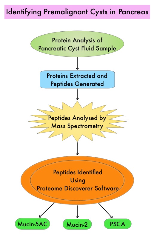
Pancreatic adenocarcinoma can also develop from mucin-producing Pancreatic Cystic Lesions (PCLs) and these neoplasms include Intraductal Papillary Mucinous Neoplasms (IPMNs) and Mucinous Cystic Neoplasms. These neoplasms comprise 10-50% of Pancreatic Cystic Lesions (PCLs). It should be noted that PCLs also encompass intrinsically benign tumors such as serous cystic neoplasms and inflammatory pseudocysts. With the rising use of abdominal MRI, partly due to concerns about ionizing radiation inherent to other exams such as CT scans, PCLs are incidentally discovered in up to 20% of these imaging studies in adults and these individuals are asymptomatic. Imaging techniques that are presently available cannot distinguish between benign, premalignant, and malignant PCLs. The same is true for currently available Endoscopic Ultrasound (EUS)-guided Fine Needle Aspiration (FNA) of Pancreatic Cystic Lesions and evaluation of cyst fluid for cytology and quantification of CarcinoEmbryonic Antigen (CEA). A high risk lesion in the pancreas would require surgical intervention with associated risks. Identifying benign from premalignant and malignant PCLs, as well as determining the epithelial subtype of IPMNs is therefore critical. The risk of malignancy is highest for Pancreatobiliary-type IPMNs with somewhat better prognosis for Intestinal-type IPMNs, whereas Gastric-type IPMNs tend to be indolent.

The authors in this study utilized targeted Mass Spectrometry (MS) to identify and quantitate proteins in the cystic fluid samples. Targeted quantitation of proteins by Mass Spectrometry provides a next-generation platform that overcomes many of the limitations of Western blotting and provides new capabilities for protein analysis. This sensitive technique is used to detect, identify and quantitate protein molecules in a given sample, based on their mass-to-charge ratio, enabling targeted protein measurement.

Using pancreatic cyst fluid samples obtained by routine EUS-guided FNA, biomarker candidates for malignant potential and high-grade dysplasia/cancer were identified via an explorative proteomic approach, in an initial cohort of 24 patients. Subsequently, a quantitative analysis using 30 heavy-labeled peptides from the biomarkers and parallel reaction monitoring mass spectrometry was devised, and tested, in a training cohort of 80 patients, and prospectively evaluated in a validation cohort of 68 patients. Patients with solid-pseudopapillary neoplasm and neuroendocrine tumor were excluded. The Primary objective of this study was to devise and validate a targeted, quantitative proteomic analysis to identify and distinguish between premalignant Pancreatic Cystic Lesions (PCLs) and Cystic neoplasms with manifest high-grade dysplasia /cancer. A Secondary aim was to find and evaluate markers for different epithelial subtypes of IPMNs, which may be used to predict the risk of malignant transformation.

It was noted that the optimal set of markers for detecting malignant potential was a panel of peptides from Mucin-5AC and Mucin-2, which could distinguish premalignant/malignant lesions from benign, with an accuracy of 97% in the validation cohort , compared with 61% using pancreatic cyst fluid CarcinoEmbryonic Antigen (P< 0.001) and 84% using Cytology (P=0.02). A combination of proteins Mucin-5AC and Prostate Stem Cell Antigen (PSCA) could identify high-grade dysplasia/cancer with an accuracy of 96% and detected 95% of malignant/severely dysplastic lesions, compared with 35% and 50% for CarcinoEmbryonic Antigen and Cytology (P<0.001 and P=0.003, respectively).

The authors concluded that Targeted Mass Spectrometry analysis of three pancreatic cyst fluid biomarkers provides highly accurate identification and assessment of cystic precursors to pancreatic adenocarcinoma. It remains to be seen whether this methodology will be beneficial for early diagnosis as well as prevention of development of pancreatic adenocarcinoma. Highly Accurate Identification of Cystic Precursor Lesions of Pancreatic Cancer Through Targeted Mass Spectrometry: A Phase IIc Diagnostic Study. Jabbar KS, Arike L, Hansson GC, et al. J Clin Oncol 2018;36:367-375

FDA Approves ZYTIGA® for High-Risk Metastatic Castration-Sensitive Prostate Cancer

SUMMARY: The FDA on February 7, 2018, approved ZYTIGA® (Abiraterone acetate) in combination with Prednisone for metastatic high-risk Castration Sensitive Prostate Cancer (CSPC). The FDA initially approved ZYTIGA® with prednisone in 2011 for patients with metastatic Castration Resistant Prostate Cancer (CRPC), who had received prior chemotherapy, and the FDA expanded the indication in 2012, for patients with chemo naïve metastatic CRPC. Prostate cancer is the most common cancer in American men with the exclusion of skin cancer and 1 in 9 men will be diagnosed with prostate cancer during their lifetime. It is estimated that in the United States, about 164,690 new cases of Prostate cancer will be diagnosed in 2018 and 29,430 men will die of the disease. The development and progression of prostate cancer is driven by androgens. Androgen Deprivation Therapy (ADT) has therefore been the cornerstone of treatment of advanced prostate cancer and is the first treatment intervention for Castration Sensitive Prostate Cancer (CSPC). Androgen Deprivation Therapies have included bilateral orchiectomy or Gonadotropin Releasing Hormone (GnRH) analogues, with or without first generation androgen receptor inhibitors such as CASODEX®: (Bicalutamide), NILANDRON® (Nilutamide) and EULEXIN® (Flutamide). The median duration of response is approximately 1 year and majority of these patients progress to Castration Resistant Prostate Cancer (CRPC). The mechanism of resistance to Androgen Deprivation Therapy (ADT) include reactivation of Androgen Receptor signaling through persistent adrenal androgen production, modification of the biologic characteristics of Androgen Receptors, intratumoral testosterone production and parallel steroidogenic pathways.Mechanism-of-Action-of-ZYTIGA

Two previously published trials, STAMPEDE and CHAARTED have shown that the addition of TAXOTERE® (Docetaxel) to Androgen Deprivaton Therapy as first line systemic therapy, significantly improved Overall Survival, among men with locally advanced or metastatic Castration Sensitive Prostate Cancer. This is presently the standard of care for appropriate patients with prostate cancer, who had not received prior hormone therapy. The barriers to chemo-hormonal therapy with TAXOTERE® include advanced patient age, poor Performance Status, comorbidities, patient preferences, as well as potential life threatening toxicities associated with TAXOTERE®.

ZYTIGA® is a selective, irreversible inhibitor of CYP 17A1 enzyme and decreases androgen biosynthesis in the testes, adrenal glands, and prostate-tumor tissue. Combining a CYP17A1 inhibitor such as ZYTIGA® with Androgen Deprivation Therapy is a more effective way of androgen depletion than with Orchiectomy or GnRH analogues alone. ZYTIGA® in combination with prednisone has been shown to significantly increase Overall Survival in patients with metastatic CRPC who had not received chemotherapy as well as those who had received previous chemotherapy with TAXOTERE®. Further, ZYTIGA® along with Prednisone has been shown to reduce tumor burden in men with high-risk, localized prostate cancer, receiving neoadjuvant therapy.

The current FDA approval was based on LATITUDE, a multinational, randomized, double-blind, placebo-controlled, phase III trial, in which the authors evaluated the clinical benefit of adding ZYTIGA® along with Prednisone to Androgen Deprivation Therapy (ADT), as compared with Androgen Deprivation Therapy and placebo, in patients with newly diagnosed, metastatic Castration Sensitive Prostate Cancer. In this study, 1199 newly diagnosed patients with high-risk metastatic prostate cancer were randomized to receive either ZYTIGA® along with Prednisone and ADT (N=597) or placebo and ADT (N=602). ZYTIGA® was administered at 1000 mg and Prednisone at 5 mg, both drugs given orally daily, and ADT consisted of a GnRH (Gonadotropin Releasing Hormone) analog. Eligible patients should not have received prior ADT and had at least 2 of 3 risk factors which included Gleason score 8 or greater, measurable visceral metastases or 3 or more bone lesions, all of which are associated with poor survival. The median age was 68 years and 98% of the enrolled patients had a Gleason score of 8 or more and had 3 or more sites of bone metastases. Both treatment groups were well balanced. The two Primary end points were Overall Survival and radiographic Progression Free Survival.

After a median follow-up of 30.4 months, at a planned interim analysis, the median Overall Survival was significantly longer in the ZYTIGA® group compared to the placebo group (Not Reached versus 34.7 months, HR=0.62; P<0.001). This meant a 38% reduction in the risk of death with the addition of ZYTIGA® and Prednisone to ADT compared with placebo and ADT. The median radiographic Progression Free Survival was 33 months in the ZYTIGA® group and 14.8 months in the placebo group (HR=0.47; P<0.001). This meant a 53% reduction in the risk of progression or death, with the addition of ZYTIGA® and Prednisone to ADT, compared with placebo and ADT. Further in the ZYTIGA® group, significantly better outcomes were observed in all Secondary end points and they included Time to pain progression, Time to next subsequent therapy for prostate cancer, Time to initiation of chemotherapy, and PSA progression (P<0.001 for all comparisons), along with Time to next symptomatic skeletal events (P=0.009). Based on these promising findings, the Independent Data and Safety Monitoring Committee recommended that the trial be unblinded and crossover be allowed for patients in the placebo group to receive ZYTIGA® and Prednisone along with ADT.

The authors concluded that the addition of ZYTIGA® along with Prednisone, to Androgen Deprivation Therapy, significantly increased Overall Survival and radiographic Progression Free Survival, in men with newly diagnosed, metastatic, Castration Sensitive Prostate Cancer. Abiraterone plus Prednisone in Metastatic, Castration-Sensitive Prostate Cancer. Fizazi K, Tran N, Fein L, et al. for the LATITUDE Investigators. N Engl J Med 2017; 377:352-360

ZYTIGA® (Abiraterone acetate)

The FDA on February 7, 2018 approved ZYTIGA® tablets in combination with Prednisone for metastatic high-risk Castration-Sensitive Prostate Cancer (CSPC). The FDA initially approved ZYTIGA® with prednisone in 2011 for patients with metastatic Castration-Resistant Prostate Cancer (CRPC), who had received prior chemotherapy, and expanded the indication in 2012 for patients with metastatic CRPC. ZYTIGA® is a product of Janssen Biotech Inc.

LUTATHERA® (Lutetium Lu 177 dotatate)

The FDA on January 26, 2018 approved LUTATHERA®, a radiolabeled Somatostatin analog, for the treatment of Somatostatin receptor-positive GastroEnteroPancreatic NeuroEndocrine Tumors (GEP-NETs), including foregut, midgut, and hindgut neuroendocrine tumors, in adults. LUTATHERA® is a product of Advanced Accelerator Applications USA, Inc.

GILOTRIF® (Afatinib)

The FDA on January 12, 2018 granted approval to GILOTRIF® for a broadened indication in first-line treatment of patients with metastatic Non-Small Cell Lung Cancer (NSCLC) whose tumors have non-resistant Epidermal Growth Factor Receptor (EGFR) mutations, as detected by an FDA-approved test. GILOTRIF® is a product of Boehringer Ingelheim Pharmaceutical, Inc.

Guideline for Human Papilloma Virus Testing in Head and Neck Carcinomas

SUMMARY: The Centers for Disease Control and Prevention estimates that in the US, there are more than 16,000 cases of Human PapillomaVirus (HPV)-positive OroPharyngeal Squamous Cell Carcinoma (OPSCC) per year and there has been a significant increase in incidence during the past several decades. They represent approximately 70% of all OPSCC in the United States and Canada. Patients with HPV-positive OPSCC tend to be younger males, who are former smokers or nonsmokers, with risk factors for exposure to High Risk HPV (HR-HPV). The HPV-positive primary Squamous Cell Carcinoma tend to be smaller in size, with early nodal metastases, and these patients have a better prognosis compared with patients with HPV-negative Head and Neck Squamous Cell Carcinoma (HNSCC), when treated similarly. Expression of tumor suppressor protein, known as p16, is highly correlated with infection with HPV in HNSCC. Accurate HPV assessment in head and neck cancers is becoming important as it significantly impacts clinical management.Molecular-Characteristics-of-HPV-Positive-Head-and-Neck-Carcinomas
There is currently no consensus on when to test oropharyngeal squamous cell carcinomas for HPV/p16, and which tests to choose. The College of American Pathologists convened a panel of experts and following review of evidence from over 400 peer reviewed articles, came up with the following Guideline. This guideline is recommended for all new Oropharyngeal Squamous cell carcinoma patients, but not routinely recommended for other head and neck carcinomas.
Summary of Guideline Statements
1) High-Risk (HR) HPV testing should be performed on all patients with newly diagnosed OPSCC, including all histologic subtypes and may be performed on the primary tumor or a regional lymph node metastasis when the clinical findings are consistent with an oropharyngeal primary. This test should not be routinely performed on nonsquamous carcinomas of the oropharynx, or nonoropharyngeal primary carcinomas of the head and neck.
2) For oropharyngeal tissue specimens (ie, noncytology), HR-HPV testing should be performed by surrogate marker p16 ImmunoHistoChemistry (IHC). Additional HPV-specific testing may be done at the discretion of the pathologist and/or treating clinician, or in the context of a clinical trial.
3) HR-HPV testing by surrogate marker p16 IHC should be routinely performed on patients with metastatic Squamous Cell Carcinoma of unknown primary in a cervical upper or mid jugular chain lymph node. An explanatory note on the significance of a positive HPV result is recommended.
4) HR-HPV testing should be performed on head and neck FNA (Fine Needle Aspiration) Squamous Cell Carcinoma samples from all patients with known OPSCC not previously tested for HR-HPV, with suspected OPSCC, or with metastatic SCC of unknown primary.
5) Pathologists should report p16 IHC positivity as a surrogate for HR-HPV in tissue specimens (ie, noncytology) when there is at least 70% nuclear and cytoplasmic expression with at least moderate to strong intensity.
6) Pathologists should not routinely perform low-risk HPV testing on patients with head and neck carcinomas.
7) For HPV-positive/p16 cases, tumor grade (or differentiation status) is not recommended.
8) HR-HPV testing strategy should not be altered based on patient smoking history.
9) Pathologists should report primary OPSCCs that test positive for HR-HPV or its surrogate marker p16 as HPV positive and/or p16 positive
Human Papillomavirus Testing in Head and Neck Carcinomas: Guideline From the College of American Pathologists. Lewis JS, Beadle B, Bishop JA, et al. https://doi.org/10.5858/arpa.2017-0286-CP

FDA Approves LUTATHERA® for the Treatment of Somatostatin Receptor-Positive GastroEnteroPancreatic Neuroendocrine Tumors

SUMMARY: The FDA on January 26, 2018 approved LUTATHERA® (Lutetium Lu 177 dotatate), a radiolabeled Somatostatin analog, for the treatment of Somatostatin receptor-positive GastroEnteroPancreatic NeuroEndocrine Tumors (GEP-NETs), including foregut, midgut, and hindgut neuroendocrine tumors, in adults. The most common type of malignant gastrointestinal NeuroEndocrine Tumors (NET) originate in the midgut (jejunoileum and the proximal colon) and often metastasize to the mesentery, peritoneum and liver. These patients frequently present with Carcinoid syndrome and are treated with Somatostatin analogue for control of tumor growth, as well as symptoms related to hormonal secretion. For patients who progress with functional neuroendocrine tumors, there are currently no standard second-line systemic treatment options available. A majority of advanced, well-differentiated neuroendocrine tumors express high levels of Somatostatin receptors and radiolabeled Somatostatin analogue therapy, also known as Peptide Receptor Radionuclide Therapy (PRRT) has been studied since the early 1990’s, with promising results. The radiolabeled Somatostatin analogues bind to the Somatostatin receptors expressed on the surface of the tumor cells and deliver targeted radiation, with a high therapeutic index, directly to the tumor cells.

LUTATHERA® is a radioconjugate consisting of the Somatostatin analog Octreotide conjugated with Lutetium-177 (177Lu), a beta and gamma-emitting radionuclide, using the chelator DOTA. In a study involving 310 patients with GastroEnteroPancreatic, NeuroEndocrine Tumors, treatment with LUTATHERA® resulted in an Objective Response Rate of 30%, and a median Progression Free Survival of 33 months.

The approval of LUTATHERA® was based on NETTER-1, a phase III, randomized, multicenter, open-label, active-controlled trial, which compared LUTATHERA® with high-dose Octreotide LAR (Long Acting), for patients with grade I or II metastatic midgut NeuroEndocrine Tumors. In this study, 229 patients (N=229) with progressive, well differentiated, locally advanced/inoperable or metastatic Somatostatin receptor-positive midgut Carcinoid tumors were randomized in a 1:1 ratio to receive either LUTATHERA® (7.4 GBq [200 mCi] every 8 weeks for up to 4 administrations along with Octreotide LAR 30 mg by IM injection every 4 weeks (N=116) or control group which received high dose Octreotide LAR 60 mg by IM injection every 4 weeks (N=113). LUTATHERA® was co-administered with an amino acid solution as a renal protectant, and in the US, patients received Aminosyn II 10%, a commercially available solution of amino acids. Well-differentiated tumors were defined as those with a Ki-67 by immunostaining of 20% or less. Tumors were assessed as low grade if they had a Ki-67 of 0-2%, intermediate grade if they had a Ki-67 of 3-20%, or high grade if they had a Ki-67 of greater than 20%, with a lower grade indicating a lower rate of cell proliferation. Baseline characteristics were well balanced between the two treatment groups. Enrolled patients had Somatostatin receptor-positive tumors and the primary site of the tumor was the ileum in 74% of the patients and the most common sites of metastasis were the liver (84%) and lymph nodes (66%). The Primary endpoint was Progression Free Survival (PFS) and Secondary endpoints included Objective Response Rates (ORR), Overall Survival (OS), and safety.

At the time of the primary analysis, it was noted that the median PFS was not reached for the LUTATHERA® group and was 8.5 months in the high-dose Octreotide LAR group (HR=0.21; P<0.001). This meant a 79% reduction in the risk of progression or death with LUTATHERA® compared with high dose Octreotide LAR. The estimated rate of PFS at month 20 was 65.2% in the LUTATHERA® group and 10.8% in the control group. The ORR with LUTATHERA® was 18% versus 3% with high dose Octreotide (P<0.001). The OS at the planned interim analysis showed a 60% reduction in the risk of death in favor of LUTATHERA® (HR=0.40; P=0.004). The most common grade 3/4 adverse reactions among patients receiving LUTATHERA® along with Octreotide LAR were nausea, vomiting, cytopenias, liver function abnormalities, hyperglycemia and hypokalemia.

It was concluded that treatment with LUTATHERA® resulted in significantly longer Progression Free Survival and a significantly higher Response Rate, when compared with high-dose Octreotide LAR, among patients with advanced midgut neuroendocrine tumors. Preliminary evidence suggests that there is an Overall Survival benefit as well. Phase 3 Trial of 177Lu-Dotatate for Midgut Neuroendocrine Tumors. Strosberg J, El-Haddad G, Wolin E, et al. for the NETTER-1 Trial Investigators. N Engl J Med 2017; 376:125-135

Molecular Testing in Lung Cancer – Guideline Update

SUMMARY: Lung cancer is the second most common cancer in both men and women and accounts for about 14% of all new cancers. The American Cancer Society estimates that for 2018 about 234,030 new cases of lung cancer will be diagnosed and over 154,050 patients will die of the disease. Non Small Cell Lung Cancer (NSCLC) accounts for approximately 85% of all lung cancers. Of the three main subtypes of Non Small Cell Lung Cancer (NSCLC), 30% are Squamous Cell Carcinomas (SCC), 40% are Adenocarcinomas and 10% are Large cell carcinomas. With changes in the cigarette composition and decline in tobacco consumption over the past several decades, Adenocarcinoma now is the most frequent histologic subtype of lung cancer.Management-of-NSCLC-based-on-Histology-and-Genomics

The College of American Pathologists, the International Association for the Study of Lung Cancer, and the Association for Molecular Pathology convened an expert panel in 2013 and had published evidence-based guideline to set standards for the molecular analysis of lung cancers and to guide treatment decisions with targeted therapies. With the availability of new medical information and technological advances, this expert panel which comprised of pathologists, oncologists, pulmonologists, and laboratory scientists, issued an evidence based update which included 18 new recommendations, along with 3 updated recommendations from the 2013 guideline, asking 5 key questions.

Key Question 1: Which new genes should be tested for lung cancer patients?

a) ROS1 testing must be performed on all lung adenocarcinoma patients, irrespective of clinical characteristics.

b) ROS1 ImmunoHistoChemistry (IHC) may be used as a screening test in lung adenocarcinoma patients; however, positive ROS1 IHC results should be confirmed by a molecular or cytogenetic method.

c) BRAF, RET, ERBB2 (HER2), KRAS and MET molecular testing are currently not indicated as a routine stand-alone assay, outside the context of a clinical trial. It is appropriate to include molecular testing for these genes, as part of larger testing panels performed either initially or when routine EGFR, ALK, and ROS1 testing are negative.

Key Question 2: What methods should be used to perform molecular testing?

a) ImmunoHistoChemistry (IHC) is an equivalent alternative to Fluorescence In Situ Hybridization (FISH) for ALK testing.

b) Multiplexed genetic sequencing panels are preferred over multiple single-gene tests, to identify other treatment options beyond EGFR, ALK, and ROS1.

c) Laboratories should ensure test results that are unexpected, discordant, equivocal or otherwise of low confidence, are confirmed or resolved, using an alternative method or sample.

Key Question 3: Is molecular testing appropriate for lung cancers that do not have an adenocarcinoma component?

a) Physicians may use molecular biomarker testing in tumors with histologies other than adenocarcinoma when clinical features indicate a higher probability of an oncogenic driver.

Key Question 4: What testing is indicated for patients with targetable mutations who have relapsed on targeted therapy?

a) In lung adenocarcinoma patients who harbor sensitizing EGFR mutations and have progressed after treatment with an EGFR-targeted TKI, physicians must use EGFR T790M mutational testing when selecting patients for third-generation EGFR-targeted therapy.

b) Laboratories testing for EGFR T790M mutation in patients with secondary clinical resistance to EGFR-targeted kinase inhibitors should deploy assays capable of detecting EGFR T790M mutations in as little as 5% of viable cells.

c) There is currently insufficient evidence to support a recommendation for or against routine testing for ALK mutational status for lung adenocarcinoma patients with sensitizing ALK mutations, who have progressed after treatment with an ALK-targeted Tyrosine Kinase Inhibitor (TKI).

Key Question 5: What is the role of testing for circulating cell-free DNA for lung cancer patients?

a) There is currently insufficient evidence to support the use of circulating cfDNA molecular methods for the diagnosis of primary lung adenocarcinoma.

b) In some clinical settings in which tissue is limited and/or insufficient for molecular testing, physicians may use a cfDNA assay to identify EGFR mutations.

c) Physicians may use cfDNA methods to identify EGFR T790M mutations in lung adenocarcinoma patients with progression or secondary clinical resistance to EGFR-targeted TKI; testing of the tumor sample is recommended if the plasma result is negative.

d) There is currently insufficient evidence to support the use of circulating tumor cell molecular analysis for the diagnosis of primary lung adenocarcinoma, the identification of EGFR or other mutations, or the identification of EGFR T790M mutations at the time of EGFR TKI resistance.

2013 Statements VERSUS 2017 Statements

a) 2013 – Cytologic samples are also suitable for EGFR and ALK testing, with cell blocks being preferred over smear preparations VERSUS 2017 – Pathologists may use either cell blocks or other cytologic preparations as suitable specimens for lung cancer biomarker molecular testing.

b) 2013 – Laboratories should use EGFR test methods that are able to detect mutations in specimens with at least 50% cancer cell content, although laboratories are strongly encouraged to use (or have available at an external reference laboratory) more sensitive tests that are able to detect mutations in specimens with as little as 10% cancer cells VERSUS 2017 – Laboratories should use, or have available at an external reference laboratory, clinical lung cancer biomarker molecular testing assays that are able to detect molecular alterations in specimens with as little as 20% cancer cells.

c) 2013 – IHC for total EGFR is not recommended for selection of EGFR TKI therapy VERSUS 2017 – It is strongly recommended that laboratories should not use total EGFR expression by IHC testing to select patients for EGFR-targeted TKI therapy.

Updated molecular testing guideline for the selection of lung cancer patients for treatment with targeted tyrosine kinase inhibitors: guideline from the College of American Pathologists, the International Association for the Study of Lung Cancer, and the Association for Molecular Pathology. Lindeman NI, Cagle PT, Aisner DL et al. [published online January 22,2018]. Arch Pathol Lab Med . doi: 10.5858/arpa.2017-0388-CP

OPDIVO® and YERVOY® Combination Improves Survival in Metastatic Colorectal Cancer

SUMMARY: ColoRectal Cancer (CRC) is the third most common cancer diagnosed in both men and women in the United States. The American Cancer Society estimates that approximately 140,250 new cases of CRC will be diagnosed in the United States in 2018 and about 50,630 patients are expected to die of the disease. The lifetime risk of developing CRC is about 1 in 21 (4.7%).

The DNA MisMatchRepair (MMR) system is responsible for molecular surveillance and works as an editing tool that identifies errors within the microsatellite regions of DNA and removes them. Defective MMR system leads to MSI (Micro Satellite Instability) and hypermutation, triggering an enhanced antitumor immune response. MSI (Micro Satellite Instability) is therefore a hallmark of defective/deficient DNA MisMatchRepair (dMMR) system and occurs in 15% of all colorectal cancers. Defective MisMatchRepair can be a sporadic or heritable event. Approximately 65% of the MSI tumors are sporadic and when sporadic, the DNA MisMatchRepair gene is MLH1. Defective MisMatchRepair can also manifest as a germline mutation occurring in 1 of the 4 MisMatchRepair genes which include MLH1, MSH2, MSH6, PMS2. This produces Lynch Syndrome (Hereditary Nonpolyposis Colorectal Carcinoma – HNPCC), an Autosomal Dominant disorder and is the most common form of hereditary colon cancer, accounting for 35% of the MSI colorectal cancers. MSI tumors tend to have better outcomes and this has been attributed to the abundance of tumor infiltrating lymphocytes in these tumors from increase immunogenicity. These tumors therefore are susceptible to PD-1 blockade with immune checkpoint inhibitors.Testing-for-MSI-and-MMR-Deficiency

MSI (Micro Satellite Instability) testing is performed using a PCR based assay and MSI-High refers to instability at 2 or more of the 5 mononucleotide repeat markers and MSI-Low refers to instability at 1 of the 5 markers. Patients are considered Micro Satellite Stable (MSS) if no instability occurs. MSI-L and MSS are grouped together because MSI-L tumors are uncommon and behave similar to MSS tumors. Tumors considered MSI-H have deficiency of one or more of the DNA MisMatchRepair genes. MMR gene deficiency can be detected by ImmunoHistoChemistry (IHC). MLH1 gene is often lost in association with PMS2. NCCN Guidelines recommend MMR or MSI testing for all patients with a history of Colon or Rectal cancer.

CheckMate 142 is a large, multi-center, open label, phase II trial which evaluated the efficacy and safety of PD-1 inhibitor treatment in patients with dMMR/MSI-H metastatic colorectal cancer. This study evaluated the benefit of OPDIVO® alone or in combination YERVOY® in this patient population. The rationale behind combining OPDIVO® a PD-1 inhibitor and YERVOY®, a CTLA-4 inhibitor, was based on the synergy between these two agents, to promote T-cell antitumor activity, thereby improving upon single-agent activity of OPDIVO®. The study enrolled 119 patients who received OPDIVO® as a single agent at 3 mg/kg IV every 2 weeks or OPDIVO® 3 mg/kg plus YERVOY® 1 mg/kg every 3 weeks for 4 doses, followed by OPDIVO® 3 mg/kg every 2 weeks. Treatment was continued until disease progression or unacceptable toxicities. The Primary endpoint was Objective Response Rate (ORR) and exploratory endpoints included Safety, Progression Free Survival (PFS), Overall Survival (OS) and efficacy in biomarker-defined populations. This study was not designed to compare the outcomes in these two treatment cohorts. Based on initial data from CheckMate-142, the FDA in July 2017 granted accelerated approval to OPDIVO® for the treatment of patients with MisMatch Repair deficient (dMMR) and MicroSatellite Instability-High (MSI-H) metastatic CRC, that has progressed, following treatment with a Fluoropyrimidine, Oxaliplatin, and Irinotecan.

This review provides an update on outcomes with monotherapy and immunotherapy combination. In the OPDIVO® monotherapy group which included 74 patients, the updated analysis at 21 months showed a response rate was 34%, with 9% being complete responses, and the disease control rate was 62%. The median duration of response has not been reached in the overall cohort of patients and among those responding, 64% had responses lasting at least 1 year. Longer follow up resulted in deepening response rates. The median Progression Free Survival (PFS) for the entire cohort was 6.6 months. Clinical Benefit was seen regardless of PD-L1 expression, BRAF mutation status, KRAS mutation status, and clinical history of Lynch Syndrome.

In the combination immunotherapy group, the median follow up was 13.4 months and the authors of this analysis, André, et al., compared the results of this cohort with those of the OPDIVO® monotherapy group, for the same 13.4 month median follow up period. The most common prior therapies included Fluoropyrimidine (99%), Oxaliplatin (93%) and Irinotecan (73%). Of the 119 patients who received this combination immunotherapy, 76% had 2 or more prior lines of therapy. The Objective Response Rate with a combination of OPDIVO® and YERVOY® was 55%, with 3.4% Complete Responses, and the Disease Control Rate was 80%. About 78% of the patients had reduction in tumor burden with combination immunotherapy. The median time to response was 2.8 months and the median Duration of Response has not yet been reached. Among patients who responded to the combination, 94% had ongoing responses at the time of data cutoff and 63% of the cohort receiving combination immunotherapy remained on treatment. These responses were noted regardless of PD-L1 expression, BRAF or KRAS mutation status, or clinical history of Lynch syndrome. The PFS and Overall Survival with combination immune checkpoint inhibitor therapy at 12 months were 71% and 85%, respectively. There were statistically significant and clinically meaningful improvements in quality-of-life measurements as well.

These data from the CheckMate-142 study support the use of OPDIVO® as a single agent or in combination with YERVOY®, for the treatment of patients with previously treated DNA MisMatch Repair-Deficient/MicroSatellite Instability-High (MSI-H) metastatic CRC.

1. Overman MJ, Bergamo F, McDermott RS, et al. Nivolumab in patients with DNA mismatch repair-deficient/microsatellite instability-high (dMMR/MSI-H) metastatic colorectal cancer (mCRC): Long-term survival according to prior line of treatment from CheckMate-142. Overman MJ, Bergamo F, McDermott RS, et al. J Clin Oncol 36, 2018 (suppl 4S; abstr 554)

2. Nivolumab + ipilimumab combination in patients with DNA mismatch repair-deficient/microsatellite instability-high (dMMR/MSI-H) metastatic colorectal cancer (mCRC): First report of the full cohort from CheckMate-142. André T, Lonardi S, Wong M, et al. J Clin Oncol 36, 2018 (suppl 4S; abstr 553)

ADCETRIS® with Chemotherapy for the Frontline Treatment of Advanced Classical Hodgkin Lymphoma

SUMMARY: The American Cancer Society estimates that in the United States for 2018, about 8,500 new cases of Hodgkin lymphoma will be diagnosed and about 1,050 patients will die of the disease. Hodgkin lymphoma is classified into two main groups – Classical Hodgkin lymphomas and Nodular Lymphocyte Predominant type, by the World Health Organization. The Classical Hodgkin lymphomas include Nodular sclerosing, Mixed cellularity, Lymphocyte rich, Lymphocyte depleted subtypes and accounts for approximately 10% of all malignant lymphomas. Nodular sclerosis Hodgkin lymphoma histology, accounts for approximately 80% of Hodgkin lymphoma cases in older children and adolescents in the United States. Classical Hodgkin Lymphoma is a malignancy of primarily B lymphocytes and is characterized by the presence of large mononucleated Hodgkin (H) and giant multinucleated Reed-Sternberg (RS) cells, collectively known as Hodgkin and Reed-Sternberg cells (HRS).WHO-Classification-of-Hodgkin-Lymphoma

For patients with Hodgkin Lymphoma, the goal of first-line chemotherapy is cure. A positive PET scan following first-line chemotherapy is indicative of incomplete response with residual disease and warrants subsequent chemotherapy or radiation. Advanced stage (stage III to stage IV) Classical Hodgkin lymphoma has a cure rate of approximately 70-80% when treated in the first-line setting with a combination of Doxorubicin, Bleomycin, Vinblastine, and Dacarbazine (ABVD). This regimen which was developed more than 40 years ago is less expensive, easy to administer, is generally well tolerated and is often used in first line setting. Nonetheless, this regimen which contains Bleomycin can cause pulmonary toxicity, the incidence of which is higher in older patients and in those who receive consolidation radiotherapy to the thorax.

ADCETRIS® (Brentuximab vedotin) is an antibody-drug conjugate (ADC) that targets CD30, which is a surface antigen, expressed on Reed-Sternberg cells, in patients with Classical Hodgkin lymphoma. This ADC consists of an anti-CD30 monoclonal antibody linked to MonoMethyl Auristatin E (MMAE), an antimicrotubule agent. Upon binding to the CD30 molecule on the cancer cells, MMAE is released into the cancer cell, resulting in cell death. ADCETRIS® is presently approved by the FDA for the treatment of Classical Hodgkin lymphoma, after failure of Autologous Hematopoietic Stem Cell Transplantation (auto-HSCT) or after failure of at least two prior multi-agent chemotherapy regimens in patients who are not auto-HSCT candidates. It is also approved for Classical Hodgkin lymphoma at high risk of relapse or progression, as auto-HSCT consolidation.

In a previously published phase I study, ADCETRIS® in combination with AVD (A+AVD) resulted in a Complete Response rate of 96% and a 5 year Overall Survival rate of 100%. Based on these finding, ECHELON-1 study was conducted, which is an international, open-label, randomized, multicenter, phase III trial, comparing A+AVD with ABVD, as frontline therapy in patients with stage III or IV Classical Hodgkin lymphoma.

This study included 1334 previously untreated patients with stage III or IV Classical Hodgkin lymphoma, who were randomly assigned in a 1:1 ratio to receive A+AVD (N=664), which consisted of ADCETRIS® 1.2 mg/kg , Doxorubicin 25 mg/m2, Vinblastine 6 mg/m2 and Dacarbazine 375 mg/m2 or ABVD (N=670), which consisted of Doxorubicin 25 mg/m2, Bleomycin 10 units/m2, Vinblastine 6 mg/m2 and Dacarbazine 375 mg/m2, given intravenously, on days 1 and 15 of each 28-day cycle, for up to 6 cycles. The Primary end point was “modified” Progression Free Survival (mPFS), which, in addition to disease progression or death, included less than Complete Response after the completion of frontline chemotherapy, based on independently assessed PET results. PET scan interpretation was based on Deauville score (The Deauville score is a 5-point scale on which higher scores indicate greater uptake of FDG glucose at involved sites on PET). Patients were stratified according to International Prognostic Score (IPS) risk group (low risk vs. intermediate risk vs. high risk). A PET scan was performed at the end of the second cycle of treatment (PET2) and patients were offered alternative frontline therapy at the discretion of the treating physician, for patients with a PET Deauville score of 5. Secondary end points included Overall Survival.

At a median follow up of 24.6 months, the 2 year modified PFS in the A+AVD and ABVD groups were 82.1% and 77.2% respectively ( HR=0.77; P=0.04). All Secondary end points also trended in favor of A+AVD. Further, the benefit of A+AVD was noted across all prespecified subgroups, including those with involvement of more than one extranodal site, patients with a high IPS risk score and stage IV disease. Additionally, a higher proportion of the patients treated with A+AVD had negative PET2 results than those treated with ABVD (89% versus 86%). There was however a higher incidence of neutropenia in the A+AVD group, but this was alleviated with G-CSF prophylaxis. There was a higher incidence of peripheral neuropathy in the A+AVD group as well, and this improved or resolved over time. Pulmonary toxicity was lower in patients receiving A+AVD compared to those receiving ABVD.

The authors concluded that at 2 years, among patients with advanced stage Hodgkin lymphoma, A+AVD had superior efficacy when compared to ABVD, with a lower combined risk of progression, death or incomplete response and subsequent use of anticancer therapy. Brentuximab Vedotin with Chemotherapy for Stage III or IV Hodgkin’s Lymphoma. Connors JM, Jurczak W, Straus DJ, et al., for the ECHELON-1 Study Group. N Engl J Med 2018; 378:331-344