GAZYVA® Superior to RITUXAN® in Follicular Lymphoma

SUMMARY: The American Cancer Society estimates that in 2017, about 72,240 people will be diagnosed with Non Hodgkin Lymphoma (NHL) in the United States and about 20,140 individuals will die of this disease. Indolent Non Hodgkin Lymphomas are mature B cell lymphoproliferative disorders and include Follicular Lymphoma, Nodal Marginal Zone Lymphoma (NMZL), Extranodal Marginal Zone Lymphoma (ENMZL) of Mucosa-Associated Lymphoid Tissue (MALT), Splenic Marginal Zone Lymphoma (SMZL), LymphoPlasmacytic Lymphoma (LPL) and Small Lymphocytic Lymphoma (SLL). Follicular Lymphoma is the most indolent form and second most common form of all NHLs and they are a heterogeneous group of lymphoproliferative malignancies. Approximately 20% of all NHLs are Follicular Lymphomas. Advanced stage indolent NHL is not curable and as such, prolonging Progression Free Survival (PFS) and Overall Survival (OS), while maintaining Quality of Life, have been the goals of treatment intervention. Asymptomatic patients with indolent NHL are generally considered candidates for “watch and wait” approach. Patients with advanced stage symptomatic Follicular Lymphoma are often treated with induction chemoimmunotherapy followed by maintenance RITUXAN® (Rituximab). This can result in a median Progression Free Survival (PFS) of 6-8 yrs and a median Overall Survival of 12-15 yrs. However, approximately 30% of the patients will relapse in 3 years.

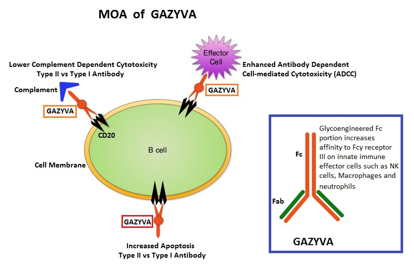
GAZYVA® (Obinutuzumab) is glycoengineered, fully humanized, third generation, type II anti-CD20 antibody (IgG1 monoclonal antibody) that selectivity binds to the extracellular domain of the CD20 antigen on malignant human B cells. By virtue of binding affinity of the glycoengineered Fc portion of GAZYVA® to Fcγ receptor III on innate immune effector cells (natural killer cells, macrophages and neutrophils), Antibody-Dependent Cell-mediated Cytotoxicity (ADCC) and Antibody-Dependent Cellular phagocytosis are significantly enhanced, but induces very little Complement-Dependent Cytotoxicity. This is in contrast to RITUXAN® which is a first generation type I, chimeric, anti-CD20 targeted monoclonal antibody that kills lymphoma cells primarily by Complement-Dependent Cytotoxicity and also ADCC.

GAZYVA® along with Bendamustine in the phase III GADOLIN study, prolonged PFS, compared with Bendamustine alone, in patients with relapsed/refractory indolent Non Hodgkin lymphoma. Based on this promising data, the GALLIUM phase III trial was conducted in treatment naïve patients with Follicular Lymphoma. This study included 1,202 patients with newly diagnosed Follicular Lymphoma, who had Grade I-IIIa tumors and had an ECOG PS of 2 or less. Patients were randomized to receive either GAZYVA® plus chemotherapy, followed by GAZYVA® maintenance (N=601), or RITUXAN® plus chemotherapy, followed by RITUXAN® maintenance (N=601). The chemotherapy regimens used were CHOP, CVP or Bendamustine, based on the discretion of the treating physician. Patients received either RITUXAN® 375mg/m2 IV on day 1 of each cycle or GAZYVA® 1000 mg IV on days 1, 8, and 15 of cycle 1 and day 1 of subsequent cycles, for either eight 21-day cycles (CHOP and CVP) or six 28-day cycles (Bendamustine). Patients who achieved a complete response (CR) or partial response (PR) at the end of induction therapy, received maintenance therapy with RITUXAN® or GAZYVA® every 2 months for 2 years or until disease progression. The median age was 59 years and 57.1% of patients received Bendamustine, 33.1% received CHOP, and 9.8% received CVP. The primary endpoint was PFS and secondary endpoints included Response Rate, Overall Survival (OS), Disease Free Survival and safety. Upon recommendations from the Independent Monitoring Committee, the study was unblinded after a preplanned interim efficacy analysis.

After a median follow-up of 34.5 months, patients in the GAZYVA® group had a 34% reduction in the risk of progression or death compared with those in the RITUXAN® group (HR=0.66; P=0.001). There was however no difference between the two treatment groups in the 3-year Overall Survival (OS) rate (P=0.21). Response rates were similar in both GAZYVA® and RITUXAN® treatment groups. Patients treated with GAZYVA® had more serious adverse events, 46.1% versus 39.9% in the RITUXAN® group, but the discontinuation rate was similar in both treatment groups.

The authors concluded that for treatment naïve Follicular Lymphoma patients, combining GAZYVA® with chemotherapy resulted in a clinically meaningful improvement in PFS compared with RITUXAN® plus chemotherapy and should therefore be the new standard of care for this patient population. Obinutuzumab-Based Induction and Maintenance Prolongs Progression-Free Survival (PFS) in Patients with Previously Untreated Follicular Lymphoma: Primary Results of the Randomized Phase 3 GALLIUM Study. Marcus RE, Davies AJ, Ando K, et al. Presented at the 58th American Society of Hematology (ASH) Annual Meeting. December 3-6, 2016; San Diego, CA. Abstract 6.

White Wine May Increase Melanoma Risk in UV-spared sites

SUMMARY: It is estimated that in the US, about 87,110 new cases of melanoma will be diagnosed in 2017 and about 9,730 patients will die of the disease. The incidence of melanoma has been on the rise for the past three decades. Alcohol related cancers account for 5.8% of all cancer deaths worldwide and there is compelling epidemiological evidence supporting that alcohol causes cancer of the oropharynx, larynx, oesophagus, liver, colon, rectum and breast (seven sites).

The mechanism of alcohol related carcinogenesis is not well understood and may vary from each target organ. Alcohol is predominantly metabolized in the liver to acetaldehyde, which is a carcinogen. Acetaldehyde is then converted into acetic acid radicals also known as acetyl radicals. There is strong evidence to suggest that acetaldehyde damages DNA. This mechanism of alcohol related carcinogenesis has been implicated in cancer of the oropharynx, larynx, esophagus and liver. With regards to breast cancer, breast tissue may be more susceptible to alcohol than other sites. Even moderate alcohol intake has been associated with increased levels of circulating sex hormones which in turn can activate cellular proliferation.

Even though the association between alcohol consumption and increased risk of numerous cancers is well known, there has been little or no evidence to associate alcohol consumption to melanoma. The authors in this study investigated whether alcohol intake was associated with risk of melanoma, by using data from three large prospective cohort studies, which included 210,252 participants. These individuals were followed for a mean of 18.3 years. The participants responded to questionnaires approximately every 4 years from 1984 to 2007 and provided information on their alcohol intake. A standard drink was defined at 12.8 grams of alcohol (one drink is considered to be 12 ounces of beer, 5 ounces of wine, or 1.5 ounces of hard liquor).

In this pooled analysis, a total of 1,374 cases of invasive melanoma were documented during the follow up period. Higher alcohol intake was associated with an increased incidence of invasive melanoma (HR=1.14; P=0.04). When analyzed by the type of alcoholic beverages and after adjusting for other alcoholic beverages, white wine consumption was associated with an increased risk of melanoma (HR 1.13; P<0.01). It was also noted that alcohol consumption-related melanoma risk, was higher in the UV-spared sites such as the torso which receives less sun exposure, compared with UV-exposed sites such as head, neck, or extremities. Individuals who consumed 20 grams or more of alcohol a day were 73% more likely to be diagnosed with melanomas of the trunk compared to nondrinkers (HR=1.3;P=0.02), whereas only 2% of the individuals were more likely to be diagnosed with melanoma of the head, neck and extremities compared with non drinkers (HR=1.02; P=0.25).

The researchers noted that there was no evidence that age, smoking history, caffeine intake, physical activity, hair color, mole count or BMI, modified the association between alcohol intake and melanoma, when these results were stratified by those variables. It was hypothesized that white wine may have higher levels of pre-existing acetaldehyde (which gives the pleasant fruity aroma), than beer or hard liquors and the antioxidants in the red wine may offset the carcinogenic risks associated with acetaldehyde.

The authors concluded that these findings support the American Cancer Society Guidelines for Cancer Prevention, to limit alcohol intake, and alcohol consumption was associated with a modest increase in the risk of melanoma, particularly on parts of the body that are less sun exposed. Alcohol Intake and Risk of Incident Melanoma: A Pooled Analysis of Three Prospective Studies in the United States. Rivera A, Nan H, Li T, et al. Cancer Epidemiol Biomarkers Prev. 2016;25:1550-1558

FDA Approves OPDIVO® for Bladder Cancer

SUMMARY: The FDA on February 2, 2017 granted accelerated approval to OPDIVO® (Nivolumab), for the treatment of patients with locally advanced or metastatic urothelial carcinoma, who have disease progression during or following platinum containing chemotherapy, or have disease progression within 12 months of neoadjuvant or adjuvant treatment with a platinum-containing chemotherapy. Urothelial carcinoma accounts for 90 percent of all bladder cancers and can originate in the renal pelvis, ureter and urethra. The American Cancer Society’s estimates that in 2017, approximately 79,030 new cases of Bladder Cancer will be diagnosed and 16,870 patients will die of the disease. Treatment options for patients who progress after platinum based chemotherapy are limited, with poor outcomes. The response rates with standard chemotherapy in this patient population, is about 10%.

The treatment paradigm for solid tumors has been rapidly evolving, with a better understanding of the Immune checkpoints or gate keepers. Immune checkpoints are cell surface inhibitory proteins/receptors that are expressed on activated T cells. They harness the immune system and prevent uncontrolled immune reactions. With the recognition of Immune checkpoint proteins and their role in suppressing antitumor immunity, antibodies are being developed that target the membrane bound inhibitory Immune checkpoint proteins/receptors such as CTLA-4 (Cytotoxic T-Lymphocyte Antigen 4, also known as CD152), PD-1(Programmed cell Death 1), as well as Programmed cell Death Ligands (PD-L1) that are expressed by cells in the tumor micro environment. By inhibiting checkpoint proteins and their ligands, T cells are unleashed, resulting in T cell proliferation, activation and a therapeutic response.

OPDIVO® is an immune checkpoint PD-1 (Programmed cell Death 1) targeted, fully human, immunoglobulin G4 monoclonal antibody that has demonstrated antitumor efficacy in multiple tumor types. The FDA approval of OPDIVO® for patients with previously treated locally advanced or metastatic urothelial carcinoma, was based on CheckMate-275 trial which is an international, multicenter, phase II study, in which 270 patients with metastatic or surgically unresectable locally advanced urothelial carcinoma received OPDIVO® 3 mg/kg IV every 2 weeks until disease progression or unacceptable toxicity. All patients had prior platinum based therapy. The median age was 66 years. The primary endpoint was overall Objective Response Rate confirmed by blinded independent review committee, in all treated patients and by tumor PD-L1 expression (5% or more and 1% or more). The follow up for this study is still ongoing. The median follow up for overall survival was 7 months.

The Objective Response Rate across all treated patients was 19.6% and the responses were durable and the median duration of response has not been reached. There was a higher likelihood of response with increasing tumor PD-L1 expression. The Objective Response Rate was 28.4% in patients with PD-L1 expression of 5% or greater, 23.8% in patients with PD-L1 expression of 1% or greater and 16.1% in those with PD-L1 expression of less than 1%. The most common adverse events were fatigue, musculoskeletal pain, nausea, and decreased appetite.

The authors concluded that single agent therapy with OPDIVO® in previously treated patients with metastatic or surgically unresectable urothelial carcinoma, resulted in durable response rate, irrespective of PD-L1 expression and was associated with an acceptable safety profile. Nivolumab in metastatic urothelial carcinoma after platinum therapy (CheckMate 275): a multicentre, single-arm, phase 2 trial. Sharma P, Retz M, Siefker-Radtke A, et al. The Lancet Oncology. Published: 25 January 2017, DOI: http://dx.doi.org/10.1016/S1470-2045(17)30065-7

The FDA recommends against using screening tests for ovarian cancer screening FDA Safety Communication

SUMMARY: The American Cancer Society estimates that about 22,440 women will be diagnosed with ovarian cancer in the United States for 2017 and about 14,080 will die of the disease. Ovarian cancer ranks fifth in cancer deaths among women, accounting for more deaths than any other cancer of the female reproductive system. Women who have reached menopause, women who have a family history of ovarian cancer, and women with the BRCA1 or BRCA2 genetic mutations have the highest risk for developing ovarian cancer. Over 75% of patients with ovarian cancer have advanced disease at the time of diagnosis. The FDA Safety Communication noted that despite extensive research and published studies there are currently no screening tests for ovarian cancer that are sensitive enough to reliably screen for ovarian cancer without a high number of inaccurate results.

The 2 tests used most often to screen for ovarian cancer are TransVaginal UltraSound (TVUS) and serum marker CA-125. TVUS is unable to differentiate benign from malignant ovarian mass. Serum marker CA-125 is usually associated with high-grade serous ovarian cancer, but is also expressed in normal tissues of the body such as the lungs and other reproductive organs. An increase in the serum marker CA-125 can be seen in non-malignant conditions such as endometriosis, peritonitis and in women with uterine fibroids. Even though serum marker CA-125 when elevated in patients, with an established diagnosis of ovarian cancer, is often used to follow the course of the disease, it has never been proven as an effective screening test for the early detection of ovarian cancer.

Nonetheless, numerous companies continue to claim that their commercially available diagnostic tests can effectively screen and detect ovarian cancer, with no data to support their claims. The FDA is concerned that women and their health care providers may rely on these inaccurate test results to make treatment decisions. Women with a false-positive result may undergo additional medical tests and/or unnecessary surgery, and may experience complications related to both. Conversely, women with a false-negative test may delay or not seek surgery or other treatment interventions for ovarian cancer. The later is particularly relevant for patients with BRCA mutations. Approximately 40% of BRCA1-mutation carriers and 18% of BRCA2-mutation carriers will develop ovarian cancer by age 70. It is recommended that patients who have BRCA1 mutations consider risk-reduction surgery (hysterectomy and bilateral bilateral salpingo-oophorectomy) by age 40 and those with BRCA2-mutations consider risk-reduction surgery no later than age 50.

Even though screenings for breast, colon and cervical cancers are successfully used for early detection and prevention of cancer-related deaths, a screening test for ovarian cancer with valid scientific data presently does not exist, and the FDA recommends against using currently offered tests to screen for ovarian cancer.

Recommendations:

For women, including those at increased risk of developing ovarian cancer

• Be aware that there is currently no safe and effective ovarian cancer screening test

• Do not rely on ovarian cancer screening test results to make health or treatment decisions

• Talk to your doctor about ways to reduce your risk of developing ovarian cancer, especially if you have a family history of ovarian cancer, or have the BRCA1 or BRCA2 genetic mutations

For physicians

• Do not recommend or use tests that claim to screen for ovarian cancer in the general population of women

• Be aware that testing higher risk asymptomatic patients for ovarian cancer has no proven benefit and is not a substitute for preventive actions that may reduce their risk

• Consider referring women at high risk of developing ovarian cancer, including those with BRCA mutations, to a genetic counselor or gynecologic oncologist, or other appropriate health care provider for more specialized care

U.S. Food and Drug Administration: The FDA recommends against using screening tests for ovarian cancer screening: FDA Safety Communication. Issued September 7, 2016. Available at http://www.fda.gov/MedicalDevices/Safety/AlertsandNotices/ucm519413.htm

OPDIVO® (Nivolumab)

The FDA on February 2, 2017 granted accelerated approval to OPDIVO® for treatment of patients with locally advanced or metastatic urothelial carcinoma who have disease progression during or following platinum-containing chemotherapy or have disease progression within 12 months of neoadjuvant or adjuvant treatment with a platinum-containing chemotherapy. OPDIVO® is marketed by Bristol-Myers Squibb company.

RUBRACA® (Rucaparib)

The FDA on November 19, 2016 granted accelerated approval to RUBRACA® for treatment of patients with deleterious BRCA mutation (germline and/or somatic) associated advanced ovarian cancer, who have been treated with two or more chemotherapies. RUBRACA® is a product of Clovis Oncology, Inc.

DARZALEX® (Daratumumab)

The FDA on November 21, 2016 approved DARZALEX® in combination with REVLIMID® (Lenalidomide) and Dexamethasone, or VELCADE® (Bortezomib) and Dexamethasone, for the treatment of patients with multiple myeloma, who have received at least one prior therapy. DARZALEX® is a product of Janssen Biotech, Inc.

OPDIVO® (Nivolumab)

The FDA on November 10, 2016 approved OPDIVO® for the treatment of patients with recurrent or metastatic Squamous Cell Carcinoma of the Head and Neck (SCCHN), with disease progression on or after a platinum-based therapy. OPDIVO® is marketed by Bristol-Myers Squibb company.

Deep Clinical Responses and MRD Negativity with DARZALEX® in Multiple Myeloma

SUMMARY: Multiple Myeloma is a clonal disorder of plasma cells in the bone marrow and the American Cancer Society estimates that in the United States, about 30,280 new cases will be diagnosed in 2017 and 12,590 patients will die of the disease. Multiple Myeloma is a disease of the elderly, with a median age at diagnosis of 69 years and characterized by intrinsic clonal heterogeneity. With a record number of regulatory approvals for Myeloma treatment over the past 12 years, the median survival for patients with Myeloma is over 10 years. The recent new drugs approved for the treatment of relapsed/refractory Multiple Myeloma include a Histone Decetylase inhibitor (FARYDAK®) and 2 monoclonal antibodies, Daratumumab (DARZALEX®) and Elotuzumab (EMPLICITI®).

Testing for MRD (Minimal Residual Disease) is standard practice in hematological malignancies such as Chronic Myeloid Leukemia (CML) which is characterized by a defined genetic abnormality ie. Philadelphia Chromosome, and CML also has very effective therapies. Unlike CML, multiple myeloma is a heterogeneous disorder and very effective therapies are only now becoming available. Effective combination regimens for myeloma has resulted in high response rates and therefore achievement of MRD negativity may be a primary endpoint in the near future. MRD negativity has been associated with prolonged Progression Free Survival (PFS) and Overall Survival (OS), in several meta-analyses.

Daratumumab (DARZALEX®) is a human IgG1 antibody that targets CD38, a transmembrane glycoprotein abundantly expressed on malignant plasma cells and with low levels of expression on normal lymphoid and myeloid cells. DARZALEX® exerts its cytotoxic effect on myeloma cells by multiple mechanisms, including Antibody Dependent Cellular Cytotoxicity (ADCC), Complement Mediated Cytotoxicity and direct apoptosis. Additionally, DARZALEX® may have a role in immunomodulation by depleting CD38-positive regulator Immune suppressor cells, and thereby expanding T cells, in patients responding to therapy.

The authors in this publication prospectively evaluated Minimal Residual Disease (MRD) status of patients enrolled in two large phase III trials, the POLLUX and CASTOR studies, and assessed the ability of DARZALEX® to yield deep clinical responses beyond complete remission. In the POLLUX study, 569 patients with relapsed or refractory multiple myeloma were randomized in a 1:1 ratio to receive either DARZALEX®, REVLIMID® (Lenalidomide) and Dexamethasone or REVLIMID® and Dexamethasone. In the CASTOR study, 498 patients with relapsed or refractory multiple myeloma were randomized in a 1:1 ratio to receive either DARZALEX®, VELCADE® (Bortezomib) and Dexamethasone or VELCADE® and Dexamethasone. In both these studies, the addition of DARZALEX® resulted in significant improvements in median PFS (HR=0.37; P<0.001 in the POLLUX study and HR=0.39; P<0.0001) in the CASTOR study), compared to the control group.

The researchers in this study, assessed MRD of bone marrow aspirate samples using ClonoSEQ next-generation sequencing–based assay. In the POLLUX study, MRD was assessed at the time of suspected Complete Response (CR), and at 3 and 6 months after. In the CASTOR study, MRD was assessed at the time of suspected CR, and at 6 months and 12 months after the first dose. The MRD sensitivity thresholds were 0.01% (1 cancer cell per 10,000 nucleated cells, or 10-4), 0.001% (10-5), and 0.0001% (10-6). The MRD negativity rate was defined as the proportion of patients with negative MRD results at any point during the studies. The median follow-up was 13.5 months for the POLLUX study and 7.4 months for the CASTOR study.

In the POLLUX study, the addition of DARZALEX® to REVLIMID® and Dexamethasone improved the MRD-negative status rates from 8.8% to 31.8% at the 10-4 threshold, from 5.7% to 24.8% at the 10-5 threshold, and from 2.5% to 11.9% at the 10-6 threshold. In the CASTOR study, the addition of DARZALEX® to VELCADE® and Dexamethasone improved the MRD-negative status rates from 3.6% to 18.3% at the 10-4 threshold, from 2.4% to 10.4% at the 10-5 threshold, and from 0.8% to 4.4% at the 10-6 threshold. It was noted that the MRD negativity was consistently higher in patients treated with DARZALEX® and DARZALEX® induced MRD negativity in three times as many patients as those treated with standard regimens. Further, MRD negativity was noted as soon as 3 months, with many patients continuing to achieve MRD negativity over time. High risk patients {t(4;14), t(14;16), del17p} benefited as well, with 18% of high risk patients in the POLLUX study and 14% of those in the CASTOR study achieving MRD negativity. Patients with sustained MRD negativity following treatment with DARZALEX®, had a significantly longer PFS compared with the control group.

It was concluded that the addition of DARZALEX®, in relapsed/refractory multiple myeloma, to standard treatment regimens, induced MRD negativity in three times as many patients as those treated with standard regimens. Evaluation of minimal residual disease (MRD) in relapsed/refractory multiple myeloma (RRMM) patients treated with daratumumab in combination with lenalidomide plus dexamethasone or bortezomib plus dexamethasone. Avet-Loisseau H, Casneuf T, Chiu C, et al. Presented at: American Society of Hematology 58th Annual Meeting; December 3-6, 2016; San Diego, CA. Abstract 246

A Tool to Predict Chemotherapy Related Toxicity in Elderly Patients

SUMMARY: Cancer in general is a disease of the elderly with two thirds of all cancers and close to three fourths of cancer mortality occurring in individuals 65 years of age or older. Elderly patients are less likely to be offered chemotherapy, as these patients are at an increased risk for chemotherapy related toxicity, compared to their younger counterparts. This may be due to a variety of reasons including decreased bone marrow reserve with aging and an increased risk of chemotherapy-related myelosuppression, inability to endure treatment due to decreased physiologic reserves, as well as impaired clearance of chemotherapy drugs due to decreased renal function. Further, cognitive and hearing impairment along with a poor support system also play an important role in the elderly, not seeking medical care, in a timely manner. Unfortunately, elderly patients are often excluded from participating in clinical trials, which makes it even more difficult to weigh the risks and benefits of chemotherapy in this patient group. Patient Performance Status is often taken into consideration to predict tolerance to chemotherapy as well as outcomes, although it remains unclear if this is a valid predictor of toxicity in elderly patients. Identifying elderly patients who are at risk for developing chemotherapy related toxicity, remains an unmet need.

In this study, researchers developed a predictive model for chemotherapy toxicity, which consisted of 11 questions pertaining to age, tumor type, treatment, lab values and geriatric assessment, using data collected from a prospective longitudinal study of 500 patients, 65 years or older. Based on the answers to the 11 questions, a score for the risk of chemotherapy toxicity was calculated and three risk groups were createdLow (0-5 points), Medium (6-9 points) and High (10-19 points). In this Development cohort, the risk for chemotherapy toxicity correlated with an increasing risk score with 53% of patients experiencing grade 3 or higher chemotherapy toxicity.

The authors then validated the predictive model in an independent cohort of elderly adults (N=250) with a median age of 73 years. It was noted that more than one half of patients in the Validation cohort (58%) experienced grade 3 or more toxicity of whom 34% experienced hematologic toxicity and 55% experienced nonhematologic toxicity. The risk of toxicity increased with increasing risk score (36.7% for low risk cohort, 62.4% for medium risk cohort and 70.2% for high risk cohort; P<0.001). These findings were not statistically different from the Development cohort. Interestingly, there was no association between Karnofsky Performance Status and chemotherapy toxicity (P=0.25).

In elderly patients receiving chemotherapy, this tool is best used to distinguish low and high risk patients but is unable to distinguish moderate and high risk patients. Further, this model was only able to predict grade 3 or more toxicity but not grade 2 toxicity, and this may be equally relevant in elderly patients. Because patients included in this study had solid tumors and did not receive biologics or high-dose chemotherapy, these results apply primarily to patients with solid tumors who receive chemotherapy.

The authors concluded that this prediction model is easy to use in clinical practice and allows Oncology Health Care Providers to help elderly patients make informed decisions and also anticipate potential toxicities in high risk elderly patients and take preventive measures accordingly. Validation of a Prediction Tool for Chemotherapy Toxicity in Older Adults with Cancer. Hurria A, Mohile S, Gajra A, et al. J Clin Oncol 2016;34:2366-2371