Circulating Tumor Cells Predict Late Recurrence in Early Stage HR-Positive Breast Cancer

SUMMARY: Breast cancer is the most common cancer among women in the US and about 1 in 8 women (12%) will develop invasive breast cancer during their lifetime. Approximately 266,120 new cases of invasive breast cancer will be diagnosed in 2018 and about 40,920 women will die of the disease. Circulating Tumor Cells (CTCs) are epithelial cells that are shed into the circulation from a primary or metastatic tumor. After being shed, CTCs can remain in the circulation or undergo apoptosis. Evaluation of CTCs during the course of disease has prognostic value. Because of the very low concentrations of CTCs (1 CTC in the background of millions of normal hematopoietic cells) in the peripheral blood, different technologies have been developed that will allow enrichment and detection of these CTCs. One such technology is the CELLSEARCH® system which is the first FDA-approved test for CTC assessment, in the peripheral blood of patients with breast cancer. This automated system is able to enrich the peripheral blood sample with CTCs and the cells then are fluorescently stained for CytoKeratins (CK8,18 and 19), Common Leukocyte Antigen (CD45) and a nuclear dye (DAPI). CTCs are identified when they are CK positive, CD45 negative and DAPI positive. In essence, CTC assessment is a real time, peripheral blood evaluation (“Liquid Biopsy”) in breast cancer patients.Differentiating-Circulating-Tumor-Cells-from-Leukocytes

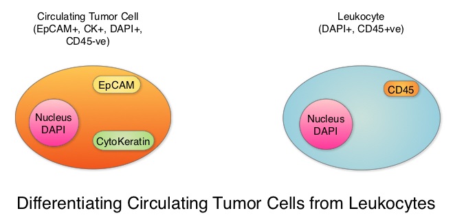
In women with Hormone Receptor (HR)-positive breast cancer, late recurrences occurring 5 or more years after diagnosis, account for approximately 50% of recurrences. In a meta-analysis from the EBCTCG (Early Breast Cancer Trialists’ Collaborative Group) group, the 10-year risk of recurrence after 5 years of endocrine therapy was found to be 5% in patients with node negative breast cancer, 10% in patients with 1-3 positive lymph nodes, and 22% in patients with 4-9 positive lymph nodes. Late recurrence in patients with HR-positive breast cancer has therefore become a major concern, as this risk for recurrence and death from breast cancer can be ongoing for at least 20 years after the original diagnosis..

The blood and tumor samples of patients are valuable for research and allow the exploration of possible tumor and host-related factors contributing to recurrence. The Coalition of Cancer Cooperative Groups, a non-profit member organization of the National Cancer Institute-sponsored Cooperative Groups, in 2013, created the North American Breast Cancer Groups (NABCG) Biospecimen Bank for Determinants of Late Relapse in Operable Breast Cancer. The purpose for the collection and analysis of samples is, to improve our understanding of breast cancer, and identify which patients are most likely to benefit from specific therapies. The NABCG Biospecimen Bank remains a unique resource of archived primary and metastatic tumor tissue, blood, and DNA, collected from more than 15,000 women, who participated in two large cooperative group cancer treatment trials, TAILORx and E5103.

The authors in this analysis included 546 patients with HER-2-negative, stage II to stage III breast cancer from the E5103 clinical trial, with no clinical evidence of recurrence between 4.5 to 7.5 years following their initial diagnosis. ECOG-ACRIN clinical trial E5103 is a randomized phase III trial, which evaluated the addition of Bevacizumab (AVASTIN®), a Vascular Endothelial Growth Factor (VEGF) inhibitor, to adjuvant chemotherapy following surgery, in patients with lymph node-positive or high-risk, lymph node-negative breast cancer. All patients had undergone surgery and adjuvant chemotherapy as well as endocrine therapy for at least 5 years, if hormone receptor positive. The CTCs from the blood samples of these patients were measured using the CELLSEARCH® CTC assay. None of the patients had clinical evidence of recurrence at the time of this analysis. More than half of the patients (56%) were 50 years or older, 59% of the patients had a tumor 2 cm or larger, 73% had positive lymph nodes and 65% had Hormone Receptor (HR) positive disease. This Primary endpoint of this study was time to recurrence and the primary focus in this analysis was on the group with Hormone Receptor positive disease.

After a median follow up of 1.6 years, 4.8% of patients had a positive CTC assay result overall (1 or more cells/7.5 mL in the blood). Among those with Hormone Receptor positive breast cancer, 5.1% had a positive CTC assay result whereas positive CTC assay result was noted in 4.1% of patients with hormone receptor negative disease. Of the 353 patients with HR-positive disease in this analysis, 4% had a recurrence whereas only 0.5% with HR-negative disease had a local recurrence. The Hazard Ratio for patients with CTC negative results compared with CTC positive results was 21.7 (P<0.001). A positive CTC assay result was associated with a nearly 21.7 fold increased risk of breast cancer recurrence in patients with HR-positive disease. A positive CTC assay was not associated with recurrence in the HR-negative group. The Positive Predictive Value of a positive CTC assay for recurrence at 2 years, in patients with HR-positive disease was 35%, and the Negative Predictive Value for patients in this cohort was 98%.

It was concluded that detection of positive Circulating Tumor Cells in the blood, 5 years after breast cancer diagnosis, was associated with an increased risk for late recurrence, in women with HR-positive, HER2-negative breast cancer. Sparano JA, O’Neill A, Alpaugh K, et al. Circulating tumor cells and late recurrence of breast cancer. Presented at: 2017 San Antonio Breast Cancer Symposium; December 5-9, 2017; San Antonio, TX. Abstract GS6-03.

Monoclonal Gammopathy of Undetermined Significance Associated with Shorter Survival

SUMMARY: Multiple Myeloma (MM) is a clonal disorder of plasma cells in the bone marrow. It evolves from a precursor stage called Monoclonal Gammopathy of Undetermined Significance (MGUS) to MM. Smoldering Multiple Myeloma (SMM) is an intermediate stage in this process of disease evolution. MGUS is defined as the presence of a serum monoclonal protein (M protein) at a concentration of 3 g/dl or less, urine with none or only modest amounts of monoclonal light chains, absence of CRAB features that are related to the M protein (Hypercalcemia, Renal insufficiency, Anemia, and Bone lesions) and 10% or fewer monoclonal plasma cells in the bone marrow. MGUS occurs in approximately 3% of individuals 50 years of age or older and in 5.3% of those 70 years of age or older.

MGUS is classified into two major subtypes, IgM and non-IgM. This is because the clonal cell that is involved and the nature of progression, differ between these two types. The origin of IgM MGUS is typically from a CD20 positive Lymphoplasmacytic cell that has not undergone Switch Recombination (Class-Switch Recombination or CSR, is a biological mechanism that changes a B cell production of immunoglobulin from one type to another, such as from the isotype IgM to the isotype IgG.), and this disease type is associated with a risk of progression to Lymphoma or Waldenström’s macroglobulinemia. In contrast, non-IgM MGUS typically arises from mature plasma cells that have undergone Switch Recombination and is associated with a risk of progression to Multiple Myeloma. Both disease types can progress to AL Amyloidosis.

There is limited information regarding malignant transformation of MGUS to Multiple Myeloma as well as Overall Survival, after diagnosis is made. Further, the risk of progression to Multiple Myeloma among the two major biologic subtypes of MGUS, IgM and non-IgM, is different, and data regarding the prognosis and risk stratification associated with these entities have not been well characterized. The authors in this publication, which is a longer follow up of previously reported findings, studied 1384 patients with MGUS, who were identified in a well-defined geographic area, from 1960-1994. The median follow up was 34.1 years. The Primary end point was progression to Multiple Myeloma or another Plasma-cell or Lymphoid disorder.

During 14,130 person-years of follow up, the rate of progression of MGUS to Multiple Myeloma was 6.5 times as high as the rate in the control population. The risk of progression to Multiple Myeloma was 10% at 10 years, 18% at 20 years, 28% at 30 years, 36% at 35 years, and 36% at 40 years. The risk of progression varied depending on the biologic subtypes of MGUS and other adverse risk factors. Among patients with IgM MGUS, the presence of two adverse risk factors – an abnormal serum free light chain ratio (ratio of kappa to lambda free light chains) and a high serum monoclonal protein level (1.5 g/dl or greater), was associated with a risk of progression at 20 years of 55%, as compared with 41% among patients who had one adverse risk factor and 19% among patients who had neither risk factor. Among patients with non-IgM MGUS, the risk of progression to Multiple Myeloma at 20 years was 30% among those who had the two risk factors, 20% among those who had one risk factor, and 7% among those who had neither risk factor. More importantly, patients with MGUS had shorter survival compared to age and sex matched control population (median 8.1 versus 12.4 years, P<0.001).

It was concluded that there were significant differences noted in the risk of progression between patients with IgM MGUS and those with non-IgM MGUS and Overall Survival was shorter among patients with MGUS than was expected in a matched control population. Long-Term Follow-up of Monoclonal Gammopathy of Undetermined Significance. Kyle RA, Larson DR, Therneau TM, et al. N Engl J Med 2018; 378:241-249

Larotrectinib – A Novel Age and Tumor Agnostic Therapy for TRK Fusion-Positive Cancers

SUMMARY: Tumor genomic profiling enables the identification of specific genomic alterations and thereby can provide personalized treatment options with targeted therapies that are specific for those molecular targets. The FDA in May 2017, granted accelerated approval to KEYTRUDA® (Pembrolizumab), for adult and pediatric patients with unresectable or metastatic, MicroSatellite Instability-High (MSI-H) or MisMatch Repair deficient (dMMR) solid tumors that have progressed following prior treatment and who have no satisfactory alternative treatment options. This is the first FDA approval of a systemic cancer treatment, based on a specific genetic biomarker, independent of tumor origin (first tissue/site-agnostic approval).

A genomic test can be performed on a tumor specimen or on cell-free DNA in plasma (“liquid biopsy”) or an ImmunoHistoChemistry (IHC) test can be performed on tumor tissue for protein expression that demonstrates a genomic variant known to be a drug target, or to predict sensitivity to a chemotherapeutic drug. Next-generation sequencing (NGS) platforms or second-generation sequencing unlike the first-generation sequencing, known as Sanger sequencing, perform massively parallel sequencing, which allows sequencing of millions of fragments of DNA from a single sample. With this high-throughput sequencing, the entire genome can be sequenced in less than 24 hours. Recently reported genomic profiling studies performed in patients with advanced cancer suggest that actionable mutations are found in 20-40% of patients’ tumors.Tropomyosin-Receptor-Kinase-Signaling-Pathway

The molecules of interest are Neurotrophic Tropomyosin Receptor Kinase (NTRK) gene rearrangements. The three TRK family of Tropomyosin Receptor Kinase (TRK) transmembrane proteins TRKA, TRKB, and TRKC are encoded by Neurotrophic Tropomyosin Receptor Kinase genes NTRK1, NTRK2, and NTRK3, respectively. These receptor tyrosine kinases are expressed in human neuronal tissue and are involved in a variety of signaling events such as cell differentiation, cell survival and apoptosis of peripheral and central neurons. They therefore play an essential role in the physiology of development and function of the nervous system. Chromosomal fusion involving NTRK genes represent the main molecular alterations with known oncogenic and transforming potential and have been identified in a variety of cancers both in children and adults. Gene fusions involving NTRK genes lead to transcription of chimeric TRK proteins which can confer oncogenic potential by increasing cell proliferation and survival. These genetic abnormalities have generated a lot of interest and have emerged as targets for cancer therapy. NGS has allowed the discovery of these gene fusions. Early clinical evidence suggests that these gene fusions lead to oncogene addiction regardless of tissue of origin. (Oncogene addiction is the dependency of some cancers on one or a few genes for the maintenance of the malignant phenotype).

Larotrectinib is a potent and highly selective small molecule inhibitor of all three TRK proteins. The authors in this development program included patients of any age and with any tumor type who had chromosomal fusion involving NTRK genes (Age and Tumor agnostic therapy). This program enrolled 55 patients, ranging in age from 4 months to 76 years, with consecutively and prospectively identified TRK fusion-positive cancers, detected by molecular profiling. They were assigned to three clinical studies – a phase I study involving adults, a phase I-II study involving children and a phase II study involving adolescents and adults.

This population of patients encompassed 17 unique cancer diagnoses and enrolled patients had locally advanced or metastatic solid tumors, and had received prior standard therapy (if available). The Primary end point for the combined analysis was the Overall Response Rate according to Independent review. Secondary end points included Duration of Response, Progression Free Survival, and safety. The researchers in this publication reported the safety and efficacy analysis of the first 55 consecutively enrolled patients, identified with TRK fusion-positive cancers, treated across these studies.

The Overall Response Rate was 75%. A total of 13% of the patients had a Complete Response, 62% had a Partial Response and 13% had stable disease. The median Duration of Response and Progression Free Survival had not been reached. At a median follow-up of 9.4 months, 86% of the patients with a response continued treatment or had undergone curative surgery. At 1 year, 71% of the responses were ongoing and 55% of the patients remained progression-free. The most common adverse events of any grade were fatigue, vomiting and abnormal liver function studies. None of the patients on Larotrectinib discontinued therapy due to a drug-related adverse event.

It was concluded that TRK fusions defined a unique molecular subgroup of advanced solid tumors in children and adults and Larotrectinib had marked and durable antitumor activity in patients with TRK fusion-positive cancer, regardless of the age of the patient or tumor type. Efficacy of Larotrectinib in TRK Fusion–Positive Cancers in Adults and Children. Drilon A, Laetsch TW, Kummar S, et al. N Engl J Med 2018; 378:731-739

Long Term Disease Free Survival Benefit with Adjuvant NERLYNX® in HER2-positive Breast Cancer

SUMMARY: Breast cancer is the most common cancer among women in the US and about 1 in 8 women (12%) will develop invasive breast cancer during their lifetime. Approximately 266,120 new cases of invasive breast cancer will be diagnosed in 2018 and about 40,920 women will die of the disease. The HER or erbB family of receptors consist of HER1, HER2, HER3 and HER4. Approximately 15-20% of invasive breast cancers overexpress HER2/neu oncogene, which is a negative predictor of outcomes without systemic therapy. HERCEPTIN® (Trastuzumab) is a humanized monoclonal antibody targeting HER2 and adjuvant chemotherapy given along with HERCEPTIN® reduces the risk of disease recurrence and death, among patients with HER2-positive, early breast cancer. Nonetheless, approximately 25% of patients will develop recurrent disease within 10 years following this adjuvant intervention. Extending the duration of adjuvant HERCEPTIN® therapy or adding TYKERB® (Lapatinib), a Tyrosine Kinase Inhibitor that targets HER1 and HER2, has not improved outcomes.HER2-Directed-Therapy

NERLYNX® is a potent, irreversible, oral Tyrosine Kinase Inhibitor, of HER1, HER2 and HER4 (pan-HER inhibitor).NERLYNX® interacts with the catalytic domain of HER1, HER2, and HER4 and blocks their downstream signaling pathways, resulting in decreased cell proliferation and increased cell death. Clinical data has suggested that NERLYNX® has significant activity in suppressing HER-mediated tumor growth and is able to overcome tumor escape mechanisms experienced with current HER2-targeted and chemotherapeutic agents. It has been well known that hormone receptor positive breast cancer patients, who are also HER2-positive, have relative resistance to hormone therapy. Preclinical models had suggested that the addition of NERLYNX® could improve responses in ER positive, HER2-positive breast cancer patients. Further, NERLYNX® has clinical activity in patients with HER2-positive metastatic breast cancer.

ExteNET trial is a multicentre, randomized, double-blind, placebo-controlled, phase III study, in which the efficacy and safety of 12 months of NERLYNX® after HERCEPTIN®-based adjuvant therapy was evaluated, in patients with early stage HER2-positive breast cancer. Patients with early stage HER2-positive breast cancer (N=2,840), and within two years of completing adjuvant HERCEPTIN®, were randomized in a 1:1 ratio to receive either oral NERLYNX® 240 mg per day (N=1420) or placebo (N=1420), for one year. Patients were stratified by hormone receptor status, nodal status (0, 1-3, or 4 or more), and HERCEPTIN® adjuvant regimen (sequentially versus concurrently with chemotherapy). The authors had previously reported that NERLYNX® when given for 12 months after chemotherapy and HERCEPTIN®-based adjuvant therapy, to women with HER2-positive breast cancer, significantly improved 2-year invasive Disease Free Survival.

They now reported updated efficacy outcomes, from a protocol-defined 5-year follow-up sensitivity analysis, and long-term toxicity findings. The predefined endpoint of the 5-year analysis was invasive Disease Free Survival (iDFS), defined as the time between the randomization date to the first occurrence of invasive recurrence (local/regional, ipsilateral or contralateral breast cancer), distant recurrence, or death from any cause. The median follow up was 5.2 years.

It was noted that patients in the NERLYNX® group had significantly fewer invasive Disease Free Survival events, than those in the placebo group (HR=0.73; P=0.0083). The 5-year invasive Disease Free Survival was 90.2% in the NERLYNX® group and 87.7% in the placebo group. The most common grade 3-4 adverse events associated with NERLYNX® were diarrhea, vomiting and nausea. Patients can experience diarrhea early, in the first 2 or 3 days and this can be alleviated using antidiarrheal prophylaxis with Loperamide, initiated with the first dose of NERLYNX® and continued for the first 2 months of treatment and as needed thereafter. There was no evidence of long-term toxicities associated with NERLYNX®, when compared to placebo.

It was concluded that at a median follow up of 5 years, NERLYNX® when given for 12 months after chemotherapy and HERCEPTIN®-based adjuvant therapy, to women with HER2-positive breast cancer, significantly reduced the proportion of clinically relevant breast cancer relapses that might lead to death, such as distant and locoregional recurrences outside the preserved breast. This benefit was seen without increase in the risk of long term toxicity. Overall Survival data will be available at a later date. NERLYNX® is the first TKI approved by the FDA, shown to reduce the risk for disease recurrence, in patients with early stage HER2-positive breast cancer. Neratinib after trastuzumab-based adjuvant therapy in HER2-positive breast cancer (ExteNET): 5-year analysis of a randomised, double-blind, placebo-controlled, phase 3 trial. Martin M, Holmes FA, Ejlertsen B, et al. for the ExteNET Study Group. Lancet Oncol 2017;18:1688-1700

Perioperative Interruption of Direct Oral Anticoagulants in Patients with Venous Thromboembolic Disease

Perioperative Interruption of Direct Oral Anticoagulants in Patients with Venous Thromboembolic Disease

SUMMARY: Direct Oral AntiCoagulants (DOACs) are often prescribed for thromboembolic events. This class of anticoagulants, have a rapid onset and offset of action, short half-life and predictable anticoagulant effects, without the need for routine monitoring. Further, several studies have demonstrated non-inferiority or superiority of this class of drugs compared with Vitamin K Aantagonists (VKAs), with regards to prevention and treatment of thromboembolic events. It is estimated that each year 10-15% of patients on DOACs will undergo an invasive procedure or surgery and will require temporary interruption of anticoagulation prior to standard-risk procedures and procedures with increased risk for bleeding. Several studies have evaluated the perioperative interruption of DOACs based on half-life of the anticoagulant and the underlying procedural bleeding risk in patient with Atrial Fibrillation. Whether these findings can be extrapolated to patients with VTE, has remained unclear.Anticoagulants-Classification

The authors in this study evaluated the thrombotic and bleeding outcomes following the perioperative interruption of Direct Oral AntiCoagulants, in patients with prior VTE (Venous ThromboEmbolism). This retrospective study included 190 patients who were on Direct Oral AntiCoagulants, such as PRADAXA® (Dabigatran), XARELTO® (Rivaroxaban), SAVAYSA® (Edoxaban) or ELIQUIS® (Apixaban), for previous VTE, and were scheduled to undergo an invasive procedure or surgery. They required temporary interruption of anticoagulation prior to standard-risk procedures and procedures with increased risk for bleeding. About 80% of the patients had unprovoked VTE, as the most recent thrombotic event, and 25% of the patients had recurrent VTE. The mean age was 59 years.Bleeding-Risk-for-Invasive-Procedures

The timing to interrupt and reinitiate Direct Oral Anticoagulant therapy was at the discretion of the treating physician, and typically DOACs were held for three half-lives prior to and restarted 2 days following standard-bleeding risk procedures, and for five half-lives prior to and restarted 4 days following high-bleeding risk procedures. The mean time from last dose of DOAC to surgery was 56.9 hours for standard-bleeding risk procedures and 69.9 hours for high-bleeding risk procedures. The mean time to therapeutic dosing of DOACs was 47 hours in the standard-bleeding risk group and 80.2 hours in the high-bleeding risk group. Approximately 41% of patients also received prophylactic doses of Low Molecular Weight Heparin or DOACs in the immediate postoperative period before re-initiation of therapeutic anticoagulation. Also taken into consideration was the elimination half-life of DOACs which can be increased by decreased renal function (PRADAXA® > SAVAYSA® > XARELTO® and ELIQUIS®), severe hepatic insufficiency (XARELTO® and ELIQUIS® > SAVAYSA® > PRADAXA®) and drug interactions. The Primary efficacy outcome was the 30-day symptomatic VTE rate, and the Primary safety outcome was the 30-day major bleeding rate. Secondary outcomes included overall mortality and the rate of clinically relevant non-major bleeding.

The 30-day VTE rate was 1.05% and the 30-day major bleeding rate was 0.53%. There were no deaths during the 30-day follow-up period. The rate of clinically relevant non-major bleeding was 3.16%.

It was concluded that perioperative interruption of Direct Oral AntiCoagulants, using a strategy that considered the half-life of the DOAC and the underlying procedural bleeding risk, appeared to be both safe and effective among patients with prior Venous ThromboEmbolism. Thrombotic and bleeding outcomes following perioperative interruption of direct oral anticoagulants in patients with venous thromboembolic disease. Shaw J, de Wit C, Le Gal G, et al. J Thromb Haemost. 2017;doi:10.1111/jth.13670

Incidental Pancreatic Cysts, Malignant Potential and Pancreatic Cancer Prevention

SUMMARY: The American Cancer Society estimates that in 2018, about 55,440 people will be diagnosed with pancreatic cancer and about 44,330 people will die of the disease. Pancreatic cancer is the fourth most common cause of cancer-related deaths in the United States and Western Europe. Unfortunately, unlike other malignancies, very little progress has been made and outcomes for patients with advanced pancreatic cancer, has been dismal. Diagnosis is often made late in the course of the disease, as patients are often asymptomatic and early tumors cannot be detected during routine physical examination. Further, precursors of pancreatic cancer evolve as microscopic lesions in the ducts and are often not visualized on imaging studies. Based on the National Cancer Institute Data Base, the 5 year observed survival rate for patients diagnosed with exocrine cancer of the Pancreas is 14% for those with Stage IA disease, and 1% for those with Stage IV disease. Early detection and cancer prevention is therefore critical.Identifying-Premalignant-Cysts-in-Pancreas 

Pancreatic adenocarcinoma can also develop from mucin-producing Pancreatic Cystic Lesions (PCLs) and these neoplasms include Intraductal Papillary Mucinous Neoplasms (IPMNs) and Mucinous Cystic Neoplasms. These neoplasms comprise 10-50% of Pancreatic Cystic Lesions (PCLs). It should be noted that PCLs also encompass intrinsically benign tumors such as serous cystic neoplasms and inflammatory pseudocysts. With the rising use of abdominal MRI, partly due to concerns about ionizing radiation inherent to other exams such as CT scans, PCLs are incidentally discovered in up to 20% of these imaging studies in adults and these individuals are asymptomatic. Imaging techniques that are presently available cannot distinguish between benign, premalignant, and malignant PCLs. The same is true for currently available Endoscopic Ultrasound (EUS)-guided Fine Needle Aspiration (FNA) of Pancreatic Cystic Lesions and evaluation of cyst fluid for cytology and quantification of CarcinoEmbryonic Antigen (CEA). A high risk lesion in the pancreas would require surgical intervention with associated risks. Identifying benign from premalignant and malignant PCLs, as well as determining the epithelial subtype of IPMNs is therefore critical. The risk of malignancy is highest for Pancreatobiliary-type IPMNs with somewhat better prognosis for Intestinal-type IPMNs, whereas Gastric-type IPMNs tend to be indolent.

The authors in this study utilized targeted Mass Spectrometry (MS) to identify and quantitate proteins in the cystic fluid samples. Targeted quantitation of proteins by Mass Spectrometry provides a next-generation platform that overcomes many of the limitations of Western blotting and provides new capabilities for protein analysis. This sensitive technique is used to detect, identify and quantitate protein molecules in a given sample, based on their mass-to-charge ratio, enabling targeted protein measurement.

Using pancreatic cyst fluid samples obtained by routine EUS-guided FNA, biomarker candidates for malignant potential and high-grade dysplasia/cancer were identified via an explorative proteomic approach, in an initial cohort of 24 patients. Subsequently, a quantitative analysis using 30 heavy-labeled peptides from the biomarkers and parallel reaction monitoring mass spectrometry was devised, and tested, in a training cohort of 80 patients, and prospectively evaluated in a validation cohort of 68 patients. Patients with solid-pseudopapillary neoplasm and neuroendocrine tumor were excluded. The Primary objective of this study was to devise and validate a targeted, quantitative proteomic analysis to identify and distinguish between premalignant Pancreatic Cystic Lesions (PCLs) and Cystic neoplasms with manifest high-grade dysplasia /cancer. A Secondary aim was to find and evaluate markers for different epithelial subtypes of IPMNs, which may be used to predict the risk of malignant transformation.

It was noted that the optimal set of markers for detecting malignant potential was a panel of peptides from Mucin-5AC and Mucin-2, which could distinguish premalignant/malignant lesions from benign, with an accuracy of 97% in the validation cohort , compared with 61% using pancreatic cyst fluid CarcinoEmbryonic Antigen (P< 0.001) and 84% using Cytology (P=0.02). A combination of proteins Mucin-5AC and Prostate Stem Cell Antigen (PSCA) could identify high-grade dysplasia/cancer with an accuracy of 96% and detected 95% of malignant/severely dysplastic lesions, compared with 35% and 50% for CarcinoEmbryonic Antigen and Cytology (P<0.001 and P=0.003, respectively).

The authors concluded that Targeted Mass Spectrometry analysis of three pancreatic cyst fluid biomarkers provides highly accurate identification and assessment of cystic precursors to pancreatic adenocarcinoma. It remains to be seen whether this methodology will be beneficial for early diagnosis as well as prevention of development of pancreatic adenocarcinoma. Highly Accurate Identification of Cystic Precursor Lesions of Pancreatic Cancer Through Targeted Mass Spectrometry: A Phase IIc Diagnostic Study. Jabbar KS, Arike L, Hansson GC, et al. J Clin Oncol 2018;36:367-375

FDA Approves ZYTIGA® for High-Risk Metastatic Castration-Sensitive Prostate Cancer

SUMMARY: The FDA on February 7, 2018, approved ZYTIGA® (Abiraterone acetate) in combination with Prednisone for metastatic high-risk Castration Sensitive Prostate Cancer (CSPC). The FDA initially approved ZYTIGA® with prednisone in 2011 for patients with metastatic Castration Resistant Prostate Cancer (CRPC), who had received prior chemotherapy, and the FDA expanded the indication in 2012, for patients with chemo naïve metastatic CRPC. Prostate cancer is the most common cancer in American men with the exclusion of skin cancer and 1 in 9 men will be diagnosed with prostate cancer during their lifetime. It is estimated that in the United States, about 164,690 new cases of Prostate cancer will be diagnosed in 2018 and 29,430 men will die of the disease. The development and progression of prostate cancer is driven by androgens. Androgen Deprivation Therapy (ADT) has therefore been the cornerstone of treatment of advanced prostate cancer and is the first treatment intervention for Castration Sensitive Prostate Cancer (CSPC). Androgen Deprivation Therapies have included bilateral orchiectomy or Gonadotropin Releasing Hormone (GnRH) analogues, with or without first generation androgen receptor inhibitors such as CASODEX®: (Bicalutamide), NILANDRON® (Nilutamide) and EULEXIN® (Flutamide). The median duration of response is approximately 1 year and majority of these patients progress to Castration Resistant Prostate Cancer (CRPC). The mechanism of resistance to Androgen Deprivation Therapy (ADT) include reactivation of Androgen Receptor signaling through persistent adrenal androgen production, modification of the biologic characteristics of Androgen Receptors, intratumoral testosterone production and parallel steroidogenic pathways.Mechanism-of-Action-of-ZYTIGA

Two previously published trials, STAMPEDE and CHAARTED have shown that the addition of TAXOTERE® (Docetaxel) to Androgen Deprivaton Therapy as first line systemic therapy, significantly improved Overall Survival, among men with locally advanced or metastatic Castration Sensitive Prostate Cancer. This is presently the standard of care for appropriate patients with prostate cancer, who had not received prior hormone therapy. The barriers to chemo-hormonal therapy with TAXOTERE® include advanced patient age, poor Performance Status, comorbidities, patient preferences, as well as potential life threatening toxicities associated with TAXOTERE®.

ZYTIGA® is a selective, irreversible inhibitor of CYP 17A1 enzyme and decreases androgen biosynthesis in the testes, adrenal glands, and prostate-tumor tissue. Combining a CYP17A1 inhibitor such as ZYTIGA® with Androgen Deprivation Therapy is a more effective way of androgen depletion than with Orchiectomy or GnRH analogues alone. ZYTIGA® in combination with prednisone has been shown to significantly increase Overall Survival in patients with metastatic CRPC who had not received chemotherapy as well as those who had received previous chemotherapy with TAXOTERE®. Further, ZYTIGA® along with Prednisone has been shown to reduce tumor burden in men with high-risk, localized prostate cancer, receiving neoadjuvant therapy.

The current FDA approval was based on LATITUDE, a multinational, randomized, double-blind, placebo-controlled, phase III trial, in which the authors evaluated the clinical benefit of adding ZYTIGA® along with Prednisone to Androgen Deprivation Therapy (ADT), as compared with Androgen Deprivation Therapy and placebo, in patients with newly diagnosed, metastatic Castration Sensitive Prostate Cancer. In this study, 1199 newly diagnosed patients with high-risk metastatic prostate cancer were randomized to receive either ZYTIGA® along with Prednisone and ADT (N=597) or placebo and ADT (N=602). ZYTIGA® was administered at 1000 mg and Prednisone at 5 mg, both drugs given orally daily, and ADT consisted of a GnRH (Gonadotropin Releasing Hormone) analog. Eligible patients should not have received prior ADT and had at least 2 of 3 risk factors which included Gleason score 8 or greater, measurable visceral metastases or 3 or more bone lesions, all of which are associated with poor survival. The median age was 68 years and 98% of the enrolled patients had a Gleason score of 8 or more and had 3 or more sites of bone metastases. Both treatment groups were well balanced. The two Primary end points were Overall Survival and radiographic Progression Free Survival.

After a median follow-up of 30.4 months, at a planned interim analysis, the median Overall Survival was significantly longer in the ZYTIGA® group compared to the placebo group (Not Reached versus 34.7 months, HR=0.62; P<0.001). This meant a 38% reduction in the risk of death with the addition of ZYTIGA® and Prednisone to ADT compared with placebo and ADT. The median radiographic Progression Free Survival was 33 months in the ZYTIGA® group and 14.8 months in the placebo group (HR=0.47; P<0.001). This meant a 53% reduction in the risk of progression or death, with the addition of ZYTIGA® and Prednisone to ADT, compared with placebo and ADT. Further in the ZYTIGA® group, significantly better outcomes were observed in all Secondary end points and they included Time to pain progression, Time to next subsequent therapy for prostate cancer, Time to initiation of chemotherapy, and PSA progression (P<0.001 for all comparisons), along with Time to next symptomatic skeletal events (P=0.009). Based on these promising findings, the Independent Data and Safety Monitoring Committee recommended that the trial be unblinded and crossover be allowed for patients in the placebo group to receive ZYTIGA® and Prednisone along with ADT.

The authors concluded that the addition of ZYTIGA® along with Prednisone, to Androgen Deprivation Therapy, significantly increased Overall Survival and radiographic Progression Free Survival, in men with newly diagnosed, metastatic, Castration Sensitive Prostate Cancer. Abiraterone plus Prednisone in Metastatic, Castration-Sensitive Prostate Cancer. Fizazi K, Tran N, Fein L, et al. for the LATITUDE Investigators. N Engl J Med 2017; 377:352-360

Guideline for Human Papilloma Virus Testing in Head and Neck Carcinomas

SUMMARY: The Centers for Disease Control and Prevention estimates that in the US, there are more than 16,000 cases of Human PapillomaVirus (HPV)-positive OroPharyngeal Squamous Cell Carcinoma (OPSCC) per year and there has been a significant increase in incidence during the past several decades. They represent approximately 70% of all OPSCC in the United States and Canada. Patients with HPV-positive OPSCC tend to be younger males, who are former smokers or nonsmokers, with risk factors for exposure to High Risk HPV (HR-HPV). The HPV-positive primary Squamous Cell Carcinoma tend to be smaller in size, with early nodal metastases, and these patients have a better prognosis compared with patients with HPV-negative Head and Neck Squamous Cell Carcinoma (HNSCC), when treated similarly. Expression of tumor suppressor protein, known as p16, is highly correlated with infection with HPV in HNSCC. Accurate HPV assessment in head and neck cancers is becoming important as it significantly impacts clinical management.Molecular-Characteristics-of-HPV-Positive-Head-and-Neck-Carcinomas
There is currently no consensus on when to test oropharyngeal squamous cell carcinomas for HPV/p16, and which tests to choose. The College of American Pathologists convened a panel of experts and following review of evidence from over 400 peer reviewed articles, came up with the following Guideline. This guideline is recommended for all new Oropharyngeal Squamous cell carcinoma patients, but not routinely recommended for other head and neck carcinomas.
Summary of Guideline Statements
1) High-Risk (HR) HPV testing should be performed on all patients with newly diagnosed OPSCC, including all histologic subtypes and may be performed on the primary tumor or a regional lymph node metastasis when the clinical findings are consistent with an oropharyngeal primary. This test should not be routinely performed on nonsquamous carcinomas of the oropharynx, or nonoropharyngeal primary carcinomas of the head and neck.
2) For oropharyngeal tissue specimens (ie, noncytology), HR-HPV testing should be performed by surrogate marker p16 ImmunoHistoChemistry (IHC). Additional HPV-specific testing may be done at the discretion of the pathologist and/or treating clinician, or in the context of a clinical trial.
3) HR-HPV testing by surrogate marker p16 IHC should be routinely performed on patients with metastatic Squamous Cell Carcinoma of unknown primary in a cervical upper or mid jugular chain lymph node. An explanatory note on the significance of a positive HPV result is recommended.
4) HR-HPV testing should be performed on head and neck FNA (Fine Needle Aspiration) Squamous Cell Carcinoma samples from all patients with known OPSCC not previously tested for HR-HPV, with suspected OPSCC, or with metastatic SCC of unknown primary.
5) Pathologists should report p16 IHC positivity as a surrogate for HR-HPV in tissue specimens (ie, noncytology) when there is at least 70% nuclear and cytoplasmic expression with at least moderate to strong intensity.
6) Pathologists should not routinely perform low-risk HPV testing on patients with head and neck carcinomas.
7) For HPV-positive/p16 cases, tumor grade (or differentiation status) is not recommended.
8) HR-HPV testing strategy should not be altered based on patient smoking history.
9) Pathologists should report primary OPSCCs that test positive for HR-HPV or its surrogate marker p16 as HPV positive and/or p16 positive
Human Papillomavirus Testing in Head and Neck Carcinomas: Guideline From the College of American Pathologists. Lewis JS, Beadle B, Bishop JA, et al. https://doi.org/10.5858/arpa.2017-0286-CP

FDA Approves LUTATHERA® for the Treatment of Somatostatin Receptor-Positive GastroEnteroPancreatic Neuroendocrine Tumors

SUMMARY: The FDA on January 26, 2018 approved LUTATHERA® (Lutetium Lu 177 dotatate), a radiolabeled Somatostatin analog, for the treatment of Somatostatin receptor-positive GastroEnteroPancreatic NeuroEndocrine Tumors (GEP-NETs), including foregut, midgut, and hindgut neuroendocrine tumors, in adults. The most common type of malignant gastrointestinal NeuroEndocrine Tumors (NET) originate in the midgut (jejunoileum and the proximal colon) and often metastasize to the mesentery, peritoneum and liver. These patients frequently present with Carcinoid syndrome and are treated with Somatostatin analogue for control of tumor growth, as well as symptoms related to hormonal secretion. For patients who progress with functional neuroendocrine tumors, there are currently no standard second-line systemic treatment options available. A majority of advanced, well-differentiated neuroendocrine tumors express high levels of Somatostatin receptors and radiolabeled Somatostatin analogue therapy, also known as Peptide Receptor Radionuclide Therapy (PRRT) has been studied since the early 1990’s, with promising results. The radiolabeled Somatostatin analogues bind to the Somatostatin receptors expressed on the surface of the tumor cells and deliver targeted radiation, with a high therapeutic index, directly to the tumor cells.

LUTATHERA® is a radioconjugate consisting of the Somatostatin analog Octreotide conjugated with Lutetium-177 (177Lu), a beta and gamma-emitting radionuclide, using the chelator DOTA. In a study involving 310 patients with GastroEnteroPancreatic, NeuroEndocrine Tumors, treatment with LUTATHERA® resulted in an Objective Response Rate of 30%, and a median Progression Free Survival of 33 months.

The approval of LUTATHERA® was based on NETTER-1, a phase III, randomized, multicenter, open-label, active-controlled trial, which compared LUTATHERA® with high-dose Octreotide LAR (Long Acting), for patients with grade I or II metastatic midgut NeuroEndocrine Tumors. In this study, 229 patients (N=229) with progressive, well differentiated, locally advanced/inoperable or metastatic Somatostatin receptor-positive midgut Carcinoid tumors were randomized in a 1:1 ratio to receive either LUTATHERA® (7.4 GBq [200 mCi] every 8 weeks for up to 4 administrations along with Octreotide LAR 30 mg by IM injection every 4 weeks (N=116) or control group which received high dose Octreotide LAR 60 mg by IM injection every 4 weeks (N=113). LUTATHERA® was co-administered with an amino acid solution as a renal protectant, and in the US, patients received Aminosyn II 10%, a commercially available solution of amino acids. Well-differentiated tumors were defined as those with a Ki-67 by immunostaining of 20% or less. Tumors were assessed as low grade if they had a Ki-67 of 0-2%, intermediate grade if they had a Ki-67 of 3-20%, or high grade if they had a Ki-67 of greater than 20%, with a lower grade indicating a lower rate of cell proliferation. Baseline characteristics were well balanced between the two treatment groups. Enrolled patients had Somatostatin receptor-positive tumors and the primary site of the tumor was the ileum in 74% of the patients and the most common sites of metastasis were the liver (84%) and lymph nodes (66%). The Primary endpoint was Progression Free Survival (PFS) and Secondary endpoints included Objective Response Rates (ORR), Overall Survival (OS), and safety.

At the time of the primary analysis, it was noted that the median PFS was not reached for the LUTATHERA® group and was 8.5 months in the high-dose Octreotide LAR group (HR=0.21; P<0.001). This meant a 79% reduction in the risk of progression or death with LUTATHERA® compared with high dose Octreotide LAR. The estimated rate of PFS at month 20 was 65.2% in the LUTATHERA® group and 10.8% in the control group. The ORR with LUTATHERA® was 18% versus 3% with high dose Octreotide (P<0.001). The OS at the planned interim analysis showed a 60% reduction in the risk of death in favor of LUTATHERA® (HR=0.40; P=0.004). The most common grade 3/4 adverse reactions among patients receiving LUTATHERA® along with Octreotide LAR were nausea, vomiting, cytopenias, liver function abnormalities, hyperglycemia and hypokalemia.

It was concluded that treatment with LUTATHERA® resulted in significantly longer Progression Free Survival and a significantly higher Response Rate, when compared with high-dose Octreotide LAR, among patients with advanced midgut neuroendocrine tumors. Preliminary evidence suggests that there is an Overall Survival benefit as well. Phase 3 Trial of 177Lu-Dotatate for Midgut Neuroendocrine Tumors. Strosberg J, El-Haddad G, Wolin E, et al. for the NETTER-1 Trial Investigators. N Engl J Med 2017; 376:125-135

Molecular Testing in Lung Cancer – Guideline Update

SUMMARY: Lung cancer is the second most common cancer in both men and women and accounts for about 14% of all new cancers. The American Cancer Society estimates that for 2018 about 234,030 new cases of lung cancer will be diagnosed and over 154,050 patients will die of the disease. Non Small Cell Lung Cancer (NSCLC) accounts for approximately 85% of all lung cancers. Of the three main subtypes of Non Small Cell Lung Cancer (NSCLC), 30% are Squamous Cell Carcinomas (SCC), 40% are Adenocarcinomas and 10% are Large cell carcinomas. With changes in the cigarette composition and decline in tobacco consumption over the past several decades, Adenocarcinoma now is the most frequent histologic subtype of lung cancer.Management-of-NSCLC-based-on-Histology-and-Genomics

The College of American Pathologists, the International Association for the Study of Lung Cancer, and the Association for Molecular Pathology convened an expert panel in 2013 and had published evidence-based guideline to set standards for the molecular analysis of lung cancers and to guide treatment decisions with targeted therapies. With the availability of new medical information and technological advances, this expert panel which comprised of pathologists, oncologists, pulmonologists, and laboratory scientists, issued an evidence based update which included 18 new recommendations, along with 3 updated recommendations from the 2013 guideline, asking 5 key questions.

Key Question 1: Which new genes should be tested for lung cancer patients?

a) ROS1 testing must be performed on all lung adenocarcinoma patients, irrespective of clinical characteristics.

b) ROS1 ImmunoHistoChemistry (IHC) may be used as a screening test in lung adenocarcinoma patients; however, positive ROS1 IHC results should be confirmed by a molecular or cytogenetic method.

c) BRAF, RET, ERBB2 (HER2), KRAS and MET molecular testing are currently not indicated as a routine stand-alone assay, outside the context of a clinical trial. It is appropriate to include molecular testing for these genes, as part of larger testing panels performed either initially or when routine EGFR, ALK, and ROS1 testing are negative.

Key Question 2: What methods should be used to perform molecular testing?

a) ImmunoHistoChemistry (IHC) is an equivalent alternative to Fluorescence In Situ Hybridization (FISH) for ALK testing.

b) Multiplexed genetic sequencing panels are preferred over multiple single-gene tests, to identify other treatment options beyond EGFR, ALK, and ROS1.

c) Laboratories should ensure test results that are unexpected, discordant, equivocal or otherwise of low confidence, are confirmed or resolved, using an alternative method or sample.

Key Question 3: Is molecular testing appropriate for lung cancers that do not have an adenocarcinoma component?

a) Physicians may use molecular biomarker testing in tumors with histologies other than adenocarcinoma when clinical features indicate a higher probability of an oncogenic driver.

Key Question 4: What testing is indicated for patients with targetable mutations who have relapsed on targeted therapy?

a) In lung adenocarcinoma patients who harbor sensitizing EGFR mutations and have progressed after treatment with an EGFR-targeted TKI, physicians must use EGFR T790M mutational testing when selecting patients for third-generation EGFR-targeted therapy.

b) Laboratories testing for EGFR T790M mutation in patients with secondary clinical resistance to EGFR-targeted kinase inhibitors should deploy assays capable of detecting EGFR T790M mutations in as little as 5% of viable cells.

c) There is currently insufficient evidence to support a recommendation for or against routine testing for ALK mutational status for lung adenocarcinoma patients with sensitizing ALK mutations, who have progressed after treatment with an ALK-targeted Tyrosine Kinase Inhibitor (TKI).

Key Question 5: What is the role of testing for circulating cell-free DNA for lung cancer patients?

a) There is currently insufficient evidence to support the use of circulating cfDNA molecular methods for the diagnosis of primary lung adenocarcinoma.

b) In some clinical settings in which tissue is limited and/or insufficient for molecular testing, physicians may use a cfDNA assay to identify EGFR mutations.

c) Physicians may use cfDNA methods to identify EGFR T790M mutations in lung adenocarcinoma patients with progression or secondary clinical resistance to EGFR-targeted TKI; testing of the tumor sample is recommended if the plasma result is negative.

d) There is currently insufficient evidence to support the use of circulating tumor cell molecular analysis for the diagnosis of primary lung adenocarcinoma, the identification of EGFR or other mutations, or the identification of EGFR T790M mutations at the time of EGFR TKI resistance.

2013 Statements VERSUS 2017 Statements

a) 2013 – Cytologic samples are also suitable for EGFR and ALK testing, with cell blocks being preferred over smear preparations VERSUS 2017 – Pathologists may use either cell blocks or other cytologic preparations as suitable specimens for lung cancer biomarker molecular testing.

b) 2013 – Laboratories should use EGFR test methods that are able to detect mutations in specimens with at least 50% cancer cell content, although laboratories are strongly encouraged to use (or have available at an external reference laboratory) more sensitive tests that are able to detect mutations in specimens with as little as 10% cancer cells VERSUS 2017 – Laboratories should use, or have available at an external reference laboratory, clinical lung cancer biomarker molecular testing assays that are able to detect molecular alterations in specimens with as little as 20% cancer cells.

c) 2013 – IHC for total EGFR is not recommended for selection of EGFR TKI therapy VERSUS 2017 – It is strongly recommended that laboratories should not use total EGFR expression by IHC testing to select patients for EGFR-targeted TKI therapy.

Updated molecular testing guideline for the selection of lung cancer patients for treatment with targeted tyrosine kinase inhibitors: guideline from the College of American Pathologists, the International Association for the Study of Lung Cancer, and the Association for Molecular Pathology. Lindeman NI, Cagle PT, Aisner DL et al. [published online January 22,2018]. Arch Pathol Lab Med . doi: 10.5858/arpa.2017-0388-CP