Sequencing Therapies in Metastatic Castrate Resistant Prostate Cancer

SUMMARY: Prostate cancer is the most common cancer in American men with the exclusion of skin cancer and 1 in 7 men will be diagnosed with prostate cancer during their lifetime. It is estimated that in the United States, about 161,360 new cases of prostate cancer will be diagnosed in 2017 and 26,730 men will die of the disease. The development and progression of prostate cancer is driven by androgens. Androgen Deprivation Therapy (ADT) has therefore been the cornerstone of treatment of advanced prostate cancer and is the first treatment intervention for hormone sensitive prostate cancer. Chemotherapy is usually considered for patients who progress on hormone therapy (Castrate Resistant Prostate Cancer-CRPC) and TAXOTERE® (Docetaxel) has been shown to improve Overall Survival (OS) of metastatic prostate cancer patients, who had progressed on Androgen Deprivation Therapy. Tumors in patients with CRPC are not androgen independent and continue to rely on Androgen Receptor signaling. Two oral Androgen Receptor Targeted Agents (ARTA) are presently available for metastatic CRPC. They include ZYTIGA® (Abiraterone) and XTANDI® (Enzalutamide). ZYTIGA® inhibits CYP 17A1 enzyme thus decreasing androgen biosynthesis and depletes adrenal and intratumoral androgens. XTANDI® competes with Testosterone and Dihydrotestosterone and avidly binds to the Androgen Receptor (AR), thereby inhibiting AR signaling and in addition inhibits translocation of the AR into the nucleus and thus inhibits the transcriptional activities of the AR. There is presently very little guidance with regards to the sequencing of these two oral agents.

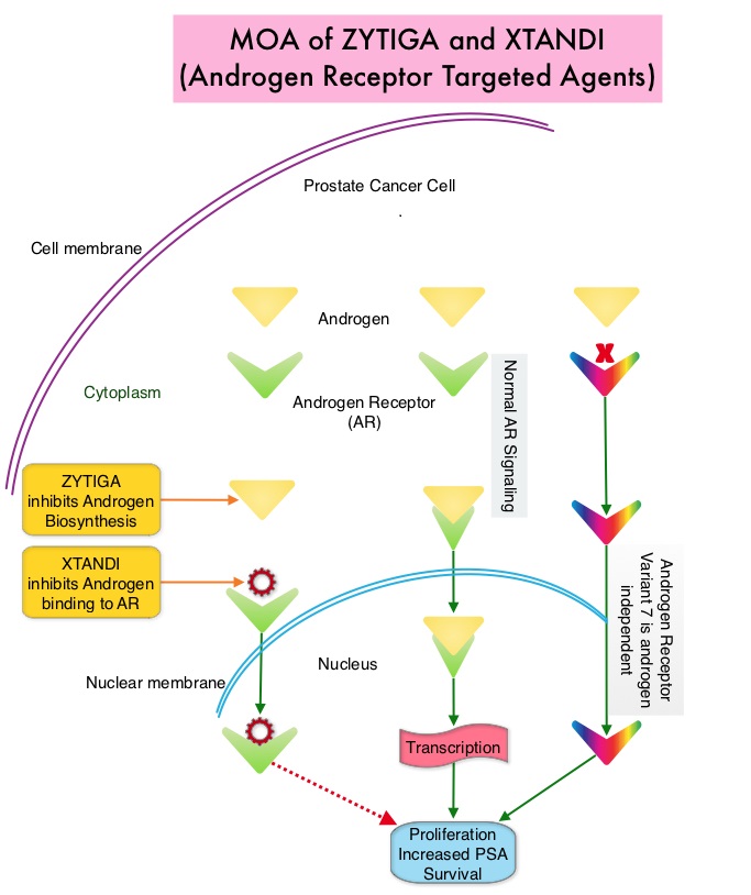
It has remained unclear when a patient should receive chemotherapy following progression on AR-targeted therapies. To determine the ideal second line therapy in this patient population, the authors conducted a retrospective study, to assess if second line chemotherapy was associated with improved outcomes, compared with second line alternative AR Targeted Agents, in patients with a very short duration of response to first line Androgen Receptor Targeted Agents, in the US community oncology setting.

Using Altos electronic medical records, the authors identified 345 patients with metastatic CRPC who did not respond to first-line AR Targeted Agents (ZYTIGA® or XTANDI®) who then went on to receive second-line TAXOTERE® – Docetaxel or JEVTANA® – Cabazitaxel (chemotherapy cohort, N=147), or an alternative AR Targeted Agent (ARTA cohort, N=198), from May 2011 to Oct 2014. Patients receiving chemotherapy as second-line treatment, compared to those receiving second-line ARTA, were younger (median age, 74 vs 79 years) and had several poor prognostic factors including a higher mean PSA, LDH and Alkaline Phosphatase, as well as lower mean hemoglobin and increased opioid use. Treatment outcomes were evaluated from start of second-line treatment and second-line chemotherapy was compared to second-line ARTA. The primary endpoints included Prostate Specific Antigen (PSA) response (50% or more reduction) and Overall Survival (OS).

It was noted that more patients in the chemotherapy group had a PSA response compared to the AR Targeted Agent group (P=0.005), and there was a non-statistically significant trend toward improved Overall Survival for second-line chemotherapy versus AR Targeted Agent (adjusted HR=0.81; P=0.15). Among patients with poor prognostic features, those in the chemotherapy cohort had significantly improved Overall Survival (adjusted HR=0.55; P=0.009) compared with those in the AR Targeted Agent cohort.

The authors concluded that patients who do not respond well to first-line Androgen Receptor Targeted Agent and have poor prognostic features, should not receive a second AR-targeted agent but instead receive second-line chemotherapy, as this may confer a survival benefit. Real-world outcomes in patients with metastatic castration-resistant prostate cancer receiving second-line chemotherapy vs alternative androgen receptor-target agents (ARTA) after lack of response to first-line ARTA in US community oncology practices. Oh WK, Cheng WY, Miao R, et al. J Clin Oncol. 2017;35 (suppl 6S; abstr 214).

Restricting Indoor Tanning Can Impact Melanoma Incidence and Treatment Costs

SUMMARY: It is estimated that in the US, about 87,110 new cases of melanoma will be diagnosed in 2017 and about 9,730 patients will die of the disease. The incidence of melanoma has been on the rise for the past three decades. A major risk factor for most skin cancers is exposure to UltraViolet (UV) radiation, which damages the DNA of skin cells. The main source of UV rays is sunlight, tanning lamps and tanning beds. UV exposure also can cause cataracts and ocular melanoma. Indoor tanning, exposes individuals to both UVA and UVB rays and is particularly dangerous for those who begin indoor tanning during adolescence or early adulthood, putting them at a higher risk of developing melanoma. The association between indoor tanning beds, including those with the newer tanning technology, and melanoma, has been well established with a 22-34% increase in the risk of developing melanoma with indoor tanning. The International Agency for Research on Cancer has classified indoor tanning as a Class I carcinogen, based on its significant association with malignant melanoma. Indoor tanning with resulting exposure to ultraviolet radiation is a potentially modifiable behavior. According to the 2015 Youth Risk Behavior Surveillance System, approximately 15% of the high school teens are indoor tanning and according to the 2010 National Health Interview Survey, indoor tanners tended to be young, non-Hispanic white women of whom 32% are aged 18 to 21 years.

The authors in this study estimated the health and economic benefits of reducing indoor tanning in the United States. Using a statistical model that takes into consideration risk, that continues over time (Markov model), the authors estimated the number of melanoma cases and deaths that could be prevented and melanoma treatment costs saved, by reducing indoor tanning among minors younger than 18 years. These estimations were based on a cohort of 61.2 million individuals aged 14 years or younger in the United States and the probability of these individuals using indoor tanning, based on data from the 2013 Youth Risk Behavior Survey. The prevalence of indoor tanning was based on a recent meta-analysis.

The researchers noted that restricting the use of indoor tanning among minors younger than 18 years was estimated to prevent 61,839 cases of melanoma (4.9% reduction) and 6,735 melanoma deaths (4.7% reduction), over the lifetime of this group of individuals. The treatment cost-savings from these reductions was estimated to be $342.9 million over the lifetime of these individuals. These health and economic benefits increased as indoor tanning was further reduced.

The authors concluded that reducing indoor tanning has the potential to reduce melanoma incidence, mortality, and treatment costs, emphasizing the importance of continued public health efforts to identify and implement effective strategies to prevent melanoma. The potential impact of reducing indoor tanning on melanoma prevention and treatment costs in the United States: An economic analysis. Guy GP, Zhang Y, Ekwueme DU, et al. Journal of the American Academy of Dermatology 2017;76:226-233

Reduced Lung-Cancer Mortality with Low-Dose Computed Tomographic Screening

SUMMARY: The rationale for Lung Cancer screening is based on the National Lung Cancer Screening Trial (NLST) in which the use of low dose CT scan in high risk, healthy patients, resulted in a 20% reduction in lung cancer mortality, compared to screening with a chest x-ray. It is important that eligible people who are smokers participate in a smoking cessation program and quit smoking. Further, those eligible for screening should understand the limitations associated with any screening methodology and potential risks associated with procedures that may follow a false positive result.

Lung cancer screening is performed using a non-contrast low dose CT scan. Criteria for lung cancer screening include-

1) People 55-74 years of age with no signs or symptoms of lung cancer

2) Current or former smoker with a 30 pack year smoking history (Number of years smoked multiplied by the number of packs of cigarettes per day)

3) Current smokers are strongly urged to enter a smoking cessation program

4) Former smokers must have quit smoking within the past 15 years

People with serious comorbid conditions, those on home oxygen and individuals with metallic devices or implants in the chest or back (which can interfere with the scan) should be excluded from lung cancer screening. Lung cancer screening with low dose CT scan is presently not covered by most insurance plans. The National Lung Screening Trial Research Team. N Engl J Med 2011; 365:395-409

CABOMETYX® Superior to SUTENT® in Metastatic Renal Cell Carcinoma

SUMMARY: The American Cancer Society estimates that about 63,990 new cases of kidney cancer will be diagnosed in the United States in 2017 and about 14,400 patients will die from this disease. The VHL (Von Hippel-Lindau) protein is a tumor suppressor gene which is frequently mutated and inactivated in approximately 90% of clear cell Renal Cell Carcinomas (RCC). The VHL gene under normal conditions binds to Hypoxia-Inducible Factor (HIF-1 alpha) and facilitates degradation of this factor. Under hypoxic conditions and in patients having biallelic loss of function and mutation of VHL genes, HIF-1alpha is not degraded. Build up of HIF-1 alpha results in increased angiogenesis, increased tumor cell proliferation and survival, as well as metastasis.

SUTENT® (Sunitinib) is the standard first-line intervention for treatment naïve patients with advanced Renal Cell Carcinoma. VOTRIENT® (Pazopanib) another VEGFR-targeted therapy, is an alternative choice, as it was found to be non-inferior to SUTENT® in the COMPARZ trial. CABOMETYX® (Cabozantinib) is an oral, small-molecule Tyrosine Kinase Inhibitor (TKI) but, unlike SUTENT® which targets the Vascular Endothelial Growth Factor Receptors (VEGFR), CABOMETYX® additionally inhibits the action of tyrosine kinases MET and AXL. Both MET and AXL are up-regulated in Renal Cell Carcinoma as a consequence of VHL inactivation and increased expression of MET and AXL is associated with tumor progression and development of resistance to VEGFR inhibitors. CABOMETYX® was approved by the FDA in 2016 for the treatment of advanced Renal Cell Carcinoma (RCC), in patients who have received prior anti-angiogenic therapy.

The Alliance for Clinical Trials in Oncology reported the results of a randomized, multicenter, open-label phase II CABOSUN trial, which compared CABOMETYX® with standard-of-care SUTENT®, in IMDC intermediate and poor-risk untreated patients with locally advanced or metastatic clear-cell RCC. The study population had a high rate of bone metastases, a known negative prognostic factor in RCC. This study enrolled 157 patients who were randomized in a 1:1 ratio to receive CABOMETYX® 60 mg orally daily (N=79) or SUTENT® 50 mg orally 4 weeks on, 2 weeks off (N=78). A treatment cycle was defined as 6 weeks in both study groups and treatment was continued until disease progression or intolerance to therapy. Patients were stratified by IMDC risk category (intermediate or poor) and presence of bone metastases. Crossover between treatment groups was not allowed. The median age was 63 years and 81% of the enrolled patients were classified as IMDC intermediate risk and 19% as poor risk, 36% of patients had bone metastases and 75% of the patients had prior nephrectomy. The Primary end point was Progression Free Survival (PFS) and Secondary end points included Objective Response Rate (ORR), Overall Survival (OS) and safety.

It was noted that the treatment with CABOMETYX® significantly increased median PFS compared with SUTENT® (8.2 versus 5.6 months) and was associated with a 34% reduction in rate of disease progression or death (HR=0.66; P=0.012). The ORR was 46% in the CABOMETYXreg; group compared with 18% in the SUTENT® group. The median OS with CABOMETYX® was 30.3 months versus 21.8 months with SUTENT® (HR=0.80), with this preliminary data showing a 20% decrease in the risk of death with CABOMETYX®. Grade 3 or 4 adverse events were similar in both treatment groups with more hypertension and Palmar-Plantar Erythrodysesthesia in the CABOMETYX® group whereas the SUTENT® group experienced more fatigue and hematologic toxicities. Discontinuation rates related to adverse events were similar in both treatment groups.

The authors concluded that CABOMETYX® significantly improved PFS and ORR compared to SUTENT®, in the initial treatment of patients with intermediate or poor-risk clear cell metastatic Renal Cell Carcinoma. CABOMETYX® is the first agent to demonstrate clinical superiority over SUTENT®, which has been the established standard of care for more than 10 years. The authors attributed the superiority of CABOMETYX® over SUTENT® due to its mechanism of action, which targets MET and AXL, in addition to VEGFR. Cabozantinib Versus Sunitinib As Initial Targeted Therapy for Patients With Metastatic Renal Cell Carcinoma of Poor or Intermediate Risk: The Alliance A031203 CABOSUN Trial. Choueiri TK, Halabi S, Sanford BL, et al. DOI: 10.1200/JCO.2016.70.7398 Journal of Clinical Oncology 35, no. 6 (February 2017) 591-597.

Location of Primary Tumor in the Colon Predicts Survival and Choice of Treatment in Metastatic Colorectal Cancer

SUMMARY: ColoRectal Cancer (CRC) is the third most common cancer diagnosed in both men and women in the United States. The American Cancer Society estimates that approximately 135,430 new cases of ColoRectal Cancer will be diagnosed in the United States in 2017 and over 50,260 patients are expected to die of the disease. Several published studies have demonstrated a nearly 20% reduced risk of death for patients diagnosed with left sided colon cancer compared with those who had right sided tumors. Venook and colleagues had previously presented their findings from a retrospective evaluation of the phase III 80405 clinical trial which included data from 1,025 metastatic ColoRectal Cancer patients with KRAS wild-type disease. The researchers assessed the impact of tumor location on Overall and Progression Free Survival in this group of patients. The median age was 59 yrs and 293 patients had a right-sided primary tumor (location in the cecum to hepatic flexure) and 732 patients had a left-sided primary tumor (location in the splenic flexure to rectum). It was noted that patients with tumors originating in the right side of the colon had much shorter median Overall Survival (19.4 months) compared to patients with left-sided tumors (33.3 months), (HR=1.60; P<0.001), with a 14 month survival improvement in the left versus right-sided primary tumors, for patients with metastatic disease. Tumor location in the colon also had a bearing on response to ERBITUX® (Cetuximab) and AVASTIN® (Bevacizumab). For the patient group who received ERBITUX®, the median Overall Survival (OS) was 36 months for patients with left-sided tumors but only 16.7 months for patients with right-sided tumors. For those who received AVASTIN®, the median OS was 31.4 months for patients with left-sided tumors and 24.2 months for those with right-sided tumors. The median Progression Free Survival was also influenced by the site of the primary tumor and was 11.5 months for left-sided tumors versus 8.9 months for right-sided tumors in the overall cohort of patients (HR = 1.26; P = 0.002). It was concluded that the biology of tumors originating in the right colon may be different from those originating in the left colon and regardless of KRAS mutational status, AVASTIN® based, first line chemotherapy regimen should be considered, for all patients with metastatic colorectal cancer, with a right-sided colon primary tumor and ERBITUX® should be avoided in this patient group.

Understanding that there is a difference in outcomes with biologic therapy, based on tumor location in the colon, the authors in this study evaluated the molecular variations of tumors arising in different parts of the left colon. Using protein expression, gene amplification and NextGen sequencing, 1,457 primary tumors (125 from splenic flexure to descending colon, 460 in the sigmoid colon and 872 in the rectum) were examined. MicroSatellite Instability (MSI) was measured by PCR and tumor mutational load was calculated using somatic nonsynonymous missense mutations.

They noted that the incidence of  to MSI significantly decreased from right colon (22%), to descending colon (7%),sigmoid colon (4%) to rectum (1%) and this was statistically significant (P=0.01). Rectal tumors had a higher frequency of TP53 and APC gene mutations and higher expression of TOPO1, ERCC1 and MGMT, compared to tumors in the descending colon. Compared to sigmoid colon tumors, rectal tumors had a higher expression of TOPO1, MGMT, TLE3 and TUBB3.

The authors concluded that tumors arising in the rectum have a distinct set of genetic alterations compared with other left colon tumors, and these distinct biologic entities may need to be addressed with individually tailored therapy. Molecular variances between rectal and left-sided colon cancers. Marshall J, Lenz H, Xiu J, et al. J Clin Oncol 35, 2017 (suppl 4S; abstract 522)

FDA Approves KEYTRUDA® for Hodgkin Lymphoma

SUMMARY: The FDA on March 14, 2017 granted an accelerated approval to KEYTRUDA® (Pembrolizumab) for the treatment of adult and pediatric patients with classical Hodgkin Lymphoma (cHL) who are Refractory, or have Relapsed, after 3 or more lines of therapy. This approved indication is based on tumor response rate and durability of response. The American Cancer Society estimates that in the United States for 2017, about 8,260 new cases of Hodgkin lymphoma will be diagnosed and about 1,070 patients will die of the disease. Hodgkin lymphoma is classified into two main groups – Classical Hodgkin lymphomas and Nodular Lymphocyte Predominant type, by the World Health Organization. The Classical Hodgkin lymphomas include Nodular sclerosing, Mixed cellularity, Lymphocyte rich, Lymphocyte depleted subtypes and accounts for approximately 10% of all malignant lymphomas. Nodular sclerosis Hodgkin lymphoma histology, accounts for approximately 80% of Hodgkin lymphoma cases in older children and adolescents in the United States. Classical Hodgkin Lymphoma is a malignancy of primarily B lymphocytes and is characterized by the presence of large mononucleated Hodgkin and giant multinucleated Reed-Sternberg (RS) cells collectively known as Hodgkin and Reed-Sternberg cells (HRS). The HRS cells in turn recruit an abundance of ineffective inflammatory cells and infiltrates of immune cells. Preclinical studies suggest that HRS cells evade immune detection by exploiting the pathways associated with immune checkpoint, Programmed Death-1 (PD-1) and its ligands PD-L. Classical Hodgkin Lymphoma is an excellent example of how the tumor microenvironment influences cancer cells to proliferate and survive.

The most common genetic abnormality in Nodular sclerosis subtype of Hodgkin lymphoma is the selective amplification of genes on the short arm of chromosome 9 (9p24.1) which includes JAK-2 with resulting increased expression of PD-1 ligands such as PDL1 and PDL2 on HRS cells, as well as increased JAK-STAT activity, essential for the proliferation and survival of Hodgkin Reed-Sternberg (HRS) cells. Infection with Epstein–Barr virus (EBV) similarly can increase the expression of PDL1 and PDL2 in EBV-positive Hodgkin’s lymphomas. It would therefore seem logical to block or inhibit immune check point PD-1 rather than both its ligands, PDL1 and PDL2. Immune checkpoints are cell surface inhibitory proteins/receptors that are expressed on activated T cells. They harness the immune system and prevent uncontrolled immune reactions. Under normal circumstances, Immune checkpoints or gate keepers inhibit intense immune responses by switching off the T cells of the immune system. With the recognition of Immune checkpoint proteins and their role in suppressing antitumor immunity, antibodies have been developed that target the membrane bound inhibitory Immune checkpoint proteins/receptors such as CTLA-4 (Cytotoxic T-Lymphocyte Antigen 4, also known as CD152), PD-1(Programmed cell Death 1), etc. By doing so, one would expect to unleash the T cells, resulting in T cell proliferation, activation and a therapeutic response. KEYTRUDA® is a fully humanized, Immunoglobulin G4, anti-PD-1, monoclonal antibody, that binds to the PD-1 receptor and blocks its interaction with ligands PD-L1 and PD-L2, thereby undoing PD-1 pathway-mediated inhibition of the immune response and unleashing the tumor-specific effector T cells.

The approval of KEYTRUDA® was based on data from the non-randomized, open-label KEYNOTE-087 trial, which included 210 adult patients with Relapsed/Refractory classical Hodgkin Lymphoma. Approximately 58% of patients were refractory to their last prior therapy, including 35% with primary refractory disease and 14% whose disease was chemo-refractory to all prior treatment regimens. Patients received a median number of four prior therapies and prior therapies included Autologous Hematopoietic Stem Cell Transplantation (61%), ADCETRIS® -Brentuximab vedotin (83%), and radiation therapy (36%). The median age was 35 years and 9% of patients were older than 65 years. KEYTRUDA® was administered at 200 mg IV every 3 weeks until disease progression, unacceptable toxicity, or for up to 2 years, in patients who did not have disease progression. Patients were assessed every 12 weeks to determine their disease status. The Primary end point was Overall Response Rate (ORR). Secondary end points included Complete Response Rate (CRR), Duration of Response, Progression Free Survival, and Overall Survival. The median follow-up was 9.4 months.

It was noted that the Overall Response Rate was 69% and this included Complete Responses in 22% of patients and Partial Responses in 47% of patients. The median Duration of Response was 11.1 months. The most common adverse events were fatigue, pyrexia, cough, musculoskeletal pain, diarrhea, and skin rash. Serious adverse reactions occurred in 16% of patients and the most common grade 3/4 treatment-related adverse events were neutropenia, thrombocytopenia and diarrhea. Adverse reactions led to treatment discontinuation in 5% of the patients.

The authors concluded that PD-1 blockade with KEYTRUDA® has significant clinical activity in subsets of heavily pretreated patients with classical Hodgkin Lymphoma, with high Overall Response Rates and Duration of Response. This important study gives a fighting chance for these generally young patients with poor prognosis. Pembrolizumab in Relapsed/Refractory Classical Hodgkin Lymphoma: Primary End Point Analysis of the Phase 2 Keynote-087 Study. Moskowitz CH, Zinzani PL, Fanale MA, et al. Presented at the ASH 58th Annual Meeting & Exposition, San Diego, CA. December 3-6, 2016. Abstract #1107

Use of Adjuvant Bisphosphonates and Other Bone-Modifying Agents in Breast Cancer A Cancer Care Ontario and American Society of Clinical Oncology Clinical Practice Guideline

SUMMARY: Breast cancer is the most common cancer among women in the US and about 1 in 8 women (12%) will develop breast cancer during their life time. Approximately, 255,180 new cases of breast cancer will be diagnosed in 2017 and 41,070 women will die of the disease. Cancer Care Ontario and ASCO convened a Working Group and Expert Panel and following a systematic review of the literature, developed evidence-based recommendations regarding the use of bisphosphonates and other bone-modifying agents, as adjuvant therapy, for patients with breast cancer. These guidelines are based on several important findings noted in previously published studies and in the more recently published Oxford Overview (Early Breast Cancer Trialists’ Collaborative Group) analysis of individual patient data.

1) Adjuvant bisphosphonates were found to reduce bone recurrence and improve survival in postmenopausal patients with non-metastatic breast cancer (including those with natural menopause or menopause induced by ovarian suppression or ablation)

2) The absolute benefit with adjuvant bisphosphonates was greater in patients who were at a higher risk of recurrence, and almost all clinical trials were conducted in patients who also received systemic therapy.

3) Most studies evaluated ZOMETA® (Zoledronic acid) or BONEFOS® (Clodronate), and there was extremely limited data for other bisphosphonates. Although XGEVA® (Denosumab) was found to reduce fractures, long-term survival data is awaited.

The following is a summary of the panel’s recommendations:

Recommendation 1

a) Administration of bisphosphonates as adjuvant therapy should be considered for postmenopausal patients with breast cancer (including patients who are premenopausal before treatment and have menopause induced by ovarian suppression as detailed in Recommendation 5) deemed candidates for adjuvant systemic therapy.

b) The final decision of whether or not to administer bisphosphonates should be made during consultation between the patient and oncologist, taking into account patient and disease characteristics, including the risk of recurrence, and weighing the potential benefits and risks.

Recommendation 2

a) Zoledronic acid and Clodronate are the recommended bisphosphonates for adjuvant therapy in breast cancer.

b) There is a need for more information comparing different agents and schedules, and it is recommended that such trials be conducted to establish the utility and optimal administration of other bisphosphonates for adjuvant therapy.

Recommendation 3

a) There is insufficient evidence at this time to make any recommendation regarding the use of Denosumab in the adjuvant setting.

b) It is recommended that studies directly comparing Denosumab with bisphosphonates and evaluating administration schedules, be conducted.

Recommendation 4

a) For patients who will receive adjuvant bisphosphonates (Recommendation 1), Zoledronic acid 4 mg IV over 15 minutes (or longer) every 6 months for 3 to 5 years or Clodronate PO 1,600 mg daily for 2 to 3 years is recommended. Different durations may be considered.

b) More research is recommended comparing different bone-modifying agents, doses, dosing intervals, and durations.

Recommendation 5

For purposes of adjuvant bisphosphonate use, the definition of menopause should include natural menopause (at least 12 months of amenorrhea prior to initiation of chemotherapy or endocrine therapy) and menopause induced by ovarian ablation or suppression (but not the cessation of menses due to chemotherapy alone). In women aged 60 years or less with a previous hysterectomy and ovaries left in place, Luteinizing Hormone, Follicle Stimulating Hormone and Serum Estradiol should be in the postmenopausal range and measured prior to initiation of any systemic therapy, to receive adjuvant bisphosphonates.

Recommendation 6

a) A dental assessment is recommended prior to commencement of bisphosphonates, where feasible, and any pending dental or oral health problems should be dealt with prior to starting treatment, if possible. Patients should be informed of the risk of developing osteonecrosis of the jaw, especially with tooth extractions and other invasive dental procedures. Patients should inform their dental practitioner of their treatment. Patients with suspected osteonecrosis of the jaw should be referred to a dental practitioner with expertise in treating this condition. Recent guidelines or position papers by groups such as the International Task Force on Osteonecrosis of the Jaw, the American Association of Oral and Maxillofacial Surgeons, and the American Dental Association should be consulted.

b) Patients should have Serum Calcium measured prior to starting treatment. Patients receiving intravenous bisphosphonates (Zoledronic acid) should be monitored for renal function, prior to starting this treatment and for Serum Calcium and increase in Serum Creatinine, throughout the treatment period.

c) Calcium and vitamin D supplementation is recommended unless otherwise contraindicated. Oral bisphosphonates and Calcium should not be taken concurrently. Several monographs suggest an interval of at least 2 hours to allow for maximum absorption.

d) Symptoms such as ocular pain or loss of vision may be due to serious inflammatory conditions such as uveitis or scleritis and should be promptly evaluated by an ophthalmologist.

Use of Adjuvant Bisphosphonates and Other Bone-Modifying Agents in Breast Cancer: A Cancer Care Ontario and American Society of Clinical Oncology Clinical Practice Guideline. Dhesy-Thind S, Fletcher GG, Blanchette PS, et al. DOI: 10.1200/JCO.2016.70.7257 Journal of Clinical Oncology – published online before print March 6, 2017

ZEJULA® (Niraparib)

The FDA on March 27, 2017 approved ZEJULA®, a poly ADP-ribose polymerase (PARP) inhibitor, for the maintenance treatment of adult patients with recurrent epithelial ovarian, fallopian tube, or primary peritoneal cancer who are in complete or partial response to platinum-based chemotherapy. ZEJULA® is marketed by Tesaro, Inc.

BAVENCIO® (Avelumab)

The FDA on March 23, 2017 granted accelerated approval to BAVENCIO® for the treatment of patients 12 years and older with metastatic Merkel Cell Carcinoma (MCC). BAVENCIO® is a programmed death-ligand 1 (PD-L1) blocking human IgG1 lambda monoclonal antibody. This is the first FDA-approved product to treat this type of cancer. BAVENCIO® is a marketed by EMD Serono, Inc.

FDA Approves KISQALI® for First-Line Treatment of Hormone Receptor Positive Advanced Breast cancer

SUMMARY: The FDA on March 13, 2017 approved KISQALI® (Ribociclib), a cyclin-dependent kinase 4/6 inhibitor, in combination with an Aromatase Inhibitor, as initial endocrine-based therapy for the treatment of postmenopausal women with Hormone Receptor (HR)-positive, Human Epidermal growth factor Receptor 2 (HER2)-negative advanced or metastatic breast cancer. Breast cancer is the most common cancer among women in the US and about 1 in 8 women (12%) will develop breast cancer during their life time. Approximately, 255,180 new cases of breast cancer will be diagnosed in 2017 and 41,070 women will die of the disease. Approximately 70% of breast tumors express Estrogen Receptors and/or Progesterone Receptors and these patients are often treated with anti-estrogen therapy as first line treatment. However, resistance to hormonal therapy occurs in a majority of the patients.

Cyclin Dependent Kinases (CDK) play a very important role to facilitate orderly and controlled progression of the cell cycle. Genetic alterations in these kinases and their regulatory proteins have been implicated in various malignancies. Cyclin Dependent Kinases 4 and 6 (CDK4 and CDK6), phosphorylate RetinoBlastoma protein (RB), and initiate transition from the G1 phase to the S phase of the cell cycle. RetinoBlastoma protein has antiproliferative and tumor-suppressor activity and phosphorylation of RB protein nullifies its beneficial activities. CDK4 and CDK6 are activated in hormone receptor positive breast cancer, promoting breast cancer cell proliferation. Further, there is evidence to suggest that endocrine resistant breast cancer cell lines depend on CDK4 for cell proliferation. The understanding of the role of Cyclin Dependent Kinases in the cell cycle, has paved the way for the development of CDK inhibitors.

KISQALI® is an orally bioavailable, selective, small-molecule inhibitor of CDK4/6 that blocks the phosphorylation of RetinoBlastoma protein, thereby preventing cell-cycle progression and inducing G1 phase arrest. In a phase 1b study involving postmenopausal women with ER positive, HER2-negative advanced breast cancer, KISQALI® in combination with FEMARA® (Letrozole) demonstrated an Overall Response Rate (ORR) of 46% and a Clinical Benefit Rate of 79%, in treatment-naïve patients with advanced breast cancer.

MONALEESA-2 trial is a randomized, double-blind, placebo-controlled, phase III study in which 668 patients were randomly assigned in a 1:1 ratio to receive either KISQALI® plus FEMARA® (Letrozole) or placebo plus FEMARA®. Eligible patients included post-menopausal women with HR-positive, HER2-negative advanced or metastatic breast cancer who had received no prior therapy for advanced disease. Treatment consisted of oral KISQALI® 600 mg daily on a 3-weeks on and 1-week off schedule, in 28-day treatment cycles plus FEMARA® 2.5 mg orally daily on a continuous schedule or placebo plus FEMARA®. Patients were stratified according to the presence or absence of liver or lung metastases and treatment was continued until disease progression or unacceptable toxicity. No treatment crossover was allowed. The median age was 62 years and close to 60% of the patients had visceral metastases. The primary end point was Progression Free Survival (PFS) and secondary end points included Overall Survival (OS), Overall Response Rate (ORR), Clinical Benefit Rate (Overall Response plus stable disease lasting 24 weeks or more), safety, and Quality of Life assessments.

A pre-planned interim efficacy analysis demonstrated a significant improvement in the PFS amongst the KISQALI® group compared to the placebo group (HR=0.56; P<0.0001). The median duration of follow-up was 15.3 months. The estimated median PFS had not been reached in the KISQALI® group and was 14.7 months in the placebo containing arm. The Overall Response Rate (ORR) in patients with measurable disease was 52.7% in the KISQALI® group and 37.1% in the placebo plus FEMARA® group (P<0.001). Overall Survival data was mature at the time of this analysis. The rates of discontinuation because of adverse events were 7.5% in the KISQALI® group and 2.1% in the placebo group. The most common adverse reactions observed in patients taking KISQALI® were myelosuppression, nausea, vomiting, diarrhea and fatigue, as well as abnormal liver function tests. KISQALI® has been shown to prolong the QT interval in a dose-dependent manner and prolongation of the QT interval occurred in 3.3% of patients treated at the 600 mg dose, with changes mostly occurring within the first 4 weeks of treatment.

The authors concluded that among patients receiving initial systemic treatment for HR-positive, HER2-negative advanced breast cancer, the addition of KISQALI® to FEMARA® significantly prolonged PFS compared to FEMARA® alone, with a higher rate of myelosuppression noted in the KISQALI® group. Ribociclib as First-Line Therapy for HR-positive, Advanced Breast Cancer. Hortobagyi GN, Stemmer SM, Burris HA, et al. N Engl J Med 375:1738-1748, 2016.