Detection of HPV DNA in the Oral Cavity Increases the Risk of Head and Neck Cancer

SUMMARY: The American Cancer Society estimates that 61,760 people will be diagnosed with Head and Neck cancer in 2016 and 13,190 patients will die of the disease. Over 90% of these malignancies are Squamous Cell Carcinomas (SCCs). Oropharyngeal Squamous Cell Carcinomas (OPSCC) involve the tonsils and base of the tongue and recent studies have shown that over 70% of these tumors are caused by Human Papilloma Virus (HPV) and HPV-16 is the predominant type present in the tumor cells. The CDC estimates that more than 2,370 new cases of Human Papilloma Virus associated Oropharyngeal Squamous Cell Carcinomas (OPSCC) are diagnosed in women and nearly 9,356 are diagnosed in men, each year in the United States and this incidence has been on the rise. The malignant behavior of these tumors is dependent on the expression of viral E6 and E7 oncoproteins that inactivate the tumor suppressor proteins p53 and the retinoblastoma protein (pRb), respectively. HPV-positive OroPharyngeal Squamous Cell Carcinoma is more common among never smokers or light smokers and patients tend to be younger with better performance status.

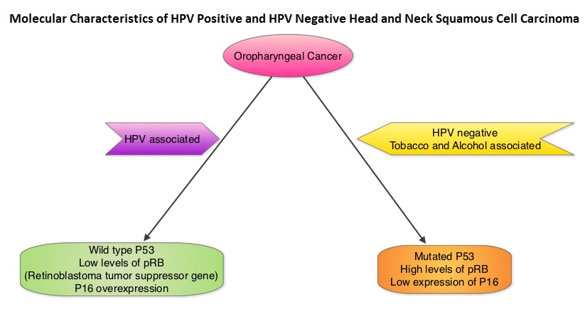
The authors conducted this prospective study to examine the temporal association between HPV DNA detection of alpha, beta and gamma Human Papilloma Virus (HPV) types in the oral samples and risk of Head and Neck Squamous Cell Carcinoma (HNSCC). A nested case-control study was carried out among 96,650 participants, cancer free at baseline, who provided mouthwash samples in 2 large prospective cohorts: the American Cancer Society Cancer Prevention Study II Nutrition Cohort and the Prostate, Lung, Colorectal, and Ovarian Cancer Screening Trial (PLCO). Molecular Detection of DNA from alpha, beta and gamma HPV types in mouthwash samples was performed by next-generation sequencing method. Associations between oral HPV infection and Head and Neck Squamous Cell Carcinoma (HNSCC) were adjusted for smoking status, pack-years and number of alcoholic drinks per week, which are all known and established risk factors for HNSCC.

During an average follow up of 3.9 years in both cohorts, 132 cases of HNSCC were identified which included cancers of the Oropharynx, Oral cavity and Larynx. The authors after analyzing the 132 cases of HNSCC and 396 controls nested within 2 prospective cohorts, found that detection of oral HPV-16 DNA was associated with a 22.4 fold increased risk of incident Oropharyngeal Cancer. Detection of oral beta1-HPV-5 type and gamma11-HPV and gamma12-HPV species was associated with a 3.3 to 5.5 fold higher risk of HNSCC.

The authors concluded that HPV-16 detection in the oral cavity precedes the incidence of Oropharyngeal Squamous Cell Carcinoma and this is the first study to demonstrate a temporal association between HPV DNA detection in mouthwash specimens and risk of HNSCC. Further, other HPVs including beta and gamma species may also play a role in the etiology of HNSCC. Detection of HPV DNA in the oral cavity may have important implications for its use in Oropharygeal cancer screening program. Associations of Oral α-, β-, and γ-Human Papillomavirus Types with Risk of Incident Head and Neck Cancer. Agalliu I, Gapstur S, Chen Z, et al. JAMA Oncol. 2016;2:599-606

XARELTO® Safe and Effective for Cancer Patients with Venous ThromboEmbolism

SUMMARY: The Center for Disease Control and Prevention (CDC) estimates that approximately 1-2 per 1000 individuals develop Deep Vein Thrombosis/Pulmonary Embolism (PE) each year in the United States, resulting in 60,000-100,000 deaths. Venous ThromboEmbolism (VTE) is the third leading cause of cardiovascular mortality. Patients with unprovoked DVT and PE are two to four times more likely to be diagnosed with cancer within the ensuing 12 months compared to the general population. Approximately 20% of VTE events are related to underlying malignancy and patients with active malignancy have a five to six fold increased risk of VTE. In patients with cancer associated thrombosis, COUMADIN® (Warfarin) and XARELTO® (Rivaroxaban) are often prescribed, despite guidelines recommending Low Molecular Weight Heparin (LMWH) in this patient population. However, patients are less inclined to take LMWH as this is parenteral, expensive, inconvenient and carries the risk of Heparin-Induced Thrombocytopenia. Further, most patients with malignancy require indefinite anticoagulation and the safety and efficacy of LMWH in this setting is unknown. The efficacy and safety of oral, direct Factor Xa inhibitor such as XARELTO® is not well established in patients with VTE and active malignancy.

The authors in this study evaluated the risk and benefits of XARELTO® in this high-risk group of patients. In this case cohort study, the Mayo Thrombophilia Clinic Direct Oral Anticoagulants Registry included patients diagnosed with Deep Vein Thrombosis or Pulmonary Embolism, who were seen and treated with XARELTO® at the Thrombophilia Clinic, Gonda Vascular Center and Mayo Clinic in Rochester, Minn. These units work together and provide streamlined standardized care. Immediate anticoagulation therapy was provided for appropriate patients. Patients with symptomatic PE or extensive symptomatic iliofemoral Deep Vein Thrombosis were hospitalized. All patients with PE also had lower extremity duplex ultrasound to determine the source of embolism. Evaluation included upper extremity venous assessment, if a patient had symptoms suggestive of venous thrombosis or if a Central Venous Catheter was present.

Patients with acute VTE or asymptomatic PE suitable for outpatient anticoagulation therapy were counseled about the pros and cons of each anticoagulant currently approved by the FDA. Additionally, patients with active malignancy and VTE were counseled about the preferred first line of treatment with Low Molecular Weight Heparin, as well as the limited data for XARELTO® in cancer-associated VTE. Patients opting for XARELTO®, were started on treatment within the ensuing hour. Patients were evaluated every 3 months for efficacy and safety and followed prospectively from March 2013 and April 2015. The primary efficacy outcome was symptomatic venous or arterial thromboembolism occurring during the follow up period. The primary safety end point was major bleeding defined as overt bleeding plus a hemoglobin decrease of 2 or more grams/dL after the incident, transfusion of 2 or more units of packed red blood cells, or intracranial, intraspinal, intraocular, retroperitoneal, pericardial, or fatal bleeding.

Two hundred and ninety six (N=296) of the 404 patients in the registry with Venous ThromboEmbolism (VTE), received XARELTO® and had at least 3 months of follow up. Of these 296 patients on XARELTO®, 118 patients (40%) had active malignancy and 178 patients had no cancer. The 3 most common cancer locations were Genitourinary (23.6%), Gastrointestinal (20.3%) and Lung (13.5%). It was noted that there was no significant difference in VTE recurrence between the malignant (3.3%) and the nonmalignant (2.8%) VTE groups (P=0.533). Slightly higher rates for major bleeding (P=0.06) and non major clinically relevant bleeding (P=0.08) were noted in patients with cancer, but this was not statistically significant.

The authors concluded that the efficacy and safety of XARELTO® is similar for VTE patients with and without active malignancy. Efficacy and Safety of Rivaroxaban in Patients with Venous Thromboembolism and Active Malignancy: A Single-Center Registry. Bott-Kitslaar DM, Saadiq RA, McBane RD, et al. Am J Med. 2016; 129: 615-619

ZYPREXA® Combination Significantly Reduces Nausea and Vomiting in Patients Receiving Highly Emetogenic Chemotherapy

SUMMARY: Chemotherapy Induced Nausea and Vomiting (CINV) is one of the most common adverse effects of chemotherapy and is experienced by about 80% of patients receiving chemotherapy. The development of effective antiemetic agents has facilitated the administration of majority of the chemotherapy agents in an outpatient setting avoiding hospitalization. Acute CINV begins within the first 24 hours following chemotherapy administration, with most patients experiencing symptoms within the first four hours of treatment, whereas delayed nausea and vomiting occurs more than 24 hours after chemotherapy administration and can persist for several days. Delayed CINV is often underestimated and a third of the patients receiving chemotherapy may experience delayed nausea and vomiting without prior acute nausea or vomiting. Acute nausea and vomiting is dependent on Serotonin (5-hydroxytryptamine-5HT3) and its receptors, with the chemotherapeutic agents stimulating the release of Serotonin from the enterochromaffin cells of the small intestine. 5-HT3 receptors are located on vagal afferent pathway, which in turn activates the vomiting center to initiate the vomiting reflex. 5-HT3 receptors are also located centrally in the Chemoreceptor Trigger Zone of the area Postrema. Delayed nausea and vomiting is associated with the activation of Neurokinin 1 (NK1) receptors by substance P. NK1 receptors are broadly distributed in the central and peripheral nervous systems.

ZYPREXA® (Olanzapine) is an antipsychotic agent that has been shown to block multiple neurotransmitters including Dopamine at D1, D2, D3, and D4 receptors, Serotonin at 5-HT2, 5-HT3 receptors, as well as Catecholamines at alpha1-adrenergic receptors, Acetylcholine at muscarinic receptors and Histamine at H1 receptors in the central nervous system. By virtue of its mechanism of action, ZYPREXA® might have significant antiemetic properties. Based on its pharmacological properties, this study evaluated the efficacy of ZYPREXA® for the prevention of nausea and vomiting, in patients receiving highly emetogenic chemotherapy.

The authors conducted a randomized, double-blind, phase III trial in which patients receiving ZYPREXA® (N=192) were compared to patients receiving Placebo (N=188). Patients received either ZYPREXA® 10 mg PO or matching Placebo daily, on days 1-4 of chemotherapy cycles. All patients additionally received Dexamethasone, Aprepitant or Fosaprepitant, and a 5-HT3 receptor antagonist. Eligible patients had no previous chemotherapy and were receiving Cisplatin at or more than 70 mg/m2 or Cyclophosphamide and Doxorubicin combination. The primary endpoint was nausea prevention and a Complete Response defined as no emesis and no use of rescue medication, was the secondary endpoint.

It was noted that the proportion of patients with no chemotherapy induced nausea was significantly greater with ZYPREXA® than with placebo in the first 24 hours after chemotherapy (74% versus. 45%, P=0.002), the time interval from 25 to 120 hours after chemotherapy (42% versus 25%, P=0.002) and across the overall 120-hour period (37% versus 22%, P=0.002). The Complete Response Rate was also significantly increased with ZYPREXA® during the three periods: 86% versus 65% (P<0.001), 67% versus 52% (P=0.007), and 64% versus 41% (P<0.001), respectively. Sedation, which is a side effect of ZYPREXA®, was observed on day 2 of treatment, with severe sedation noted in 5% of the patients.

The authors concluded that ZYPREXA® significantly improved nausea prevention, as well as Complete Response rate when compared to placebo, among previously untreated patients receiving highly emetogenic chemotherapy. Olanzapine for the Prevention of Chemotherapy-Induced Nausea and Vomiting. Navari RM, Qin R, Ruddy KJ, et al. N Engl J Med 2016; 375:134-142

Impressive 5-Year Survival Rates for Patients with Metastatic Melanoma Treated with OPDIVO®

SUMMARY: It is estimated that in the US, approximately 76,380 new cases of melanoma will be diagnosed in 2016 and approximately 10,130 patients will die of the disease. The incidence of melanoma has been on the rise for the past three decades. A better understanding of Immune checkpoints has opened the doors for the development of various immunotherapies. Immune checkpoints are cell surface inhibitory proteins/receptors that harness the immune system and prevent uncontrolled immune reactions. Survival of cancer cells in the human body may be related to their ability to escape immune surveillance, by inhibiting T lymphocyte activation. Under normal circumstances, inhibition of an intense immune response and switching off the T cells of the immune system is accomplished by Immune checkpoints or gate keepers. With the recognition of Immune checkpoint proteins and their role in suppressing antitumor immunity, antibodies have been developed that target the membrane bound inhibitory Immune checkpoint proteins/receptors such as CTLA-4 (Cytotoxic T-Lymphocyte Antigen 4, also known as CD152), PD-1(Programmed cell Death 1), etc. By blocking the Immune checkpoint proteins, one would expect to unleash the T cells, resulting in T cell proliferation, activation and a therapeutic response.

OPDIVO® (Nivolumab) is a fully human, immunoglobulin G4 monoclonal antibody that targets PD-1 receptor. In 2014, Topalian and Colleagues reported their finding of an early phase I trial (J Clin Oncol 2014;32:1020-1030), in which patients with advanced Melanoma (N = 107) who had 1-5 prior systemic therapies received OPDIVO® monotherapy. The median age of patients in this study was 61 years and patients received OPDIVO® every 2 weeks for up to 96 weeks. The median Overall Survival for these patients was 16.8 months and 1 and 2 year survival rates were 62% and 43%, respectively. It was noted that some patients had durable responses that persisted even after treatment was discontinued. This patient group was followed for Overall Survival, Progression Free Survival (PFS), long term safety and response duration, after discontinuing treatment with OPDIVO®. The authors have now reported the results of this extended follow up, with 5 year Overall Survival (OS) data from this study.

The 5 year Overall Survival in all 107 patients was 34% and the median OS was 20.3 months for those who received the approved dose of 3 mg/kg of OPDIVO® and 17.3 months in all 107 patients. As a comparison, according to the National Cancer Institute’s Surveillance, Epidemiology, and End Results (SEER) data, the 5 year OS rate for metastatic Melanoma patients diagnosed during the study period was 16.6%. The OS rates in this study appeared to have plateaued at 48 months. The Progression Free Survival at 30 months following treatment was 25.7% for those who received the approved dose of 3 mg/kg of OPDIVO® and 18.6% for all patients. The most common side effects across the entire cohort included fatigue, rash, diarrhea, pruritus and nausea.

The authors concluded that this analysis represents the first and longest follow up to date, testing an anti–PD1 immunotherapy in any specific disease. Monotherapy with OPDIVO® in heavily pretreated advanced Melanoma patients can result in more than a third of patients (34%) being alive, 5 years after starting treatment. Durable, long-term survival in previously treated patients with advanced melanoma (MEL) who received nivolumab (NIVO) monotherapy in a phase I trial. Hodi FS, Kluger H, Sznol M, et al. 2016 AACR Annual Meeting. Abstract CT001

Short Course Neoadjuvant Radiation Therapy Effective and Less Toxic in Advanced Rectal Cancer

SUMMARY: The American Cancer Society estimates 39,220 new cases of Rectal cancer will be diagnosed in the United States in 2016. Rectal cancer diagnosed at an early stage such as Stage II (T3-T4, N0) or Stage III (Node positive disease without distant metastases) is potentially curable with a combination of neoadjuvant (preoperative) chemoradiation, surgery and postoperative chemotherapy. Unlike colon cancer, the risk of locoregional recurrence is high in Rectal cancer due to its close proximity to the surrounding pelvic organs and difficulty in obtaining a clear surgical margins. Further, there is no serosal tissue surrounding the rectum. For all these reasons, preoperative Radiation Therapy (RT) with concurrent Fluoropyrimidine based chemotherapy as a radiosensitizer, followed by postoperative chemotherapy, has been the standard intervention. Radiation consists of 45 Gy delivered in 25 fractions 5 days a week with a 5.4 Gy boost. Concurrent chemotherapy in the US has included 5-FU/Leucovorin, single agent 5-FU or single agent XELODA® (Capecitabine).

The authors in this study evaluated the efficacy of a short course of neoadjuvant radiation therapy for patients with unresectable cT3 or cT4 Rectal adenocarcinoma. The trial included 515 patients who were randomly assigned either to the control group (N=254), in which patients received RT at 50.4 Gy delivered in 28 fractions, given simultaneously with a regimen of 5-FU bolus, Leucovorin and ELOXATIN® (Oxaliplatin) or the experimental group (N=261) in which patients received a short Five day course of Radiotherapy at 5 Gy per day (Total 25 Gy), followed by three courses of FOLFOX4 delivered over 48 hours, during weeks 3, 5, and 7. Both treatment groups underwent surgery approximately 12 weeks after radiation was started and about 6 weeks following neoadjuvant treatment. ELOXATIN® inclusion in the chemotherapy regimen was at the discretion of the treating physician, due to increase in toxicity. Nonetheless, 70% of the patients had received ELOXATIN® at the end of the study. It should be noted that the NSABP protocol R-04 demonstrated that the addition of ELOXATIN® in the neoadjuvant chemotherapy regimen did not improve outcomes but resulted in significant toxicity. The median follow up was 35 months.

The primary endpoint of the rate of curative resection (R0) was 71% in the control group versus 77% in the experimental group. Pathological Complete Response rates were 11.5% in the control group and 16% in the experimental group. The 3 year Overall Survival rates for the control versus experimental group were 65% vs 73% and Disease Free Survival for the control versus experimental group were 52% vs 53%. These differences were not statistically significant. The local failure rates were similar in both treatment groups (22%).

The authors concluded that a short course of radiotherapy combined with three cycles of chemotherapy post radiation, can be more convenient with lower toxicity and this regimen would be an appealing option for patients with locally advanced Rectal cancer, with metastases in liver or lungs, for whom chemotherapy to control systemic disease can be started much earlier, following a short course of radiation therapy. Neoadjuvant chemoradiation for fixed cT3 or cT4 rectal cancer: Results of a Polish II multicentre phase III study. Bujko K, et al. J Clin Oncol 34, 2016 (suppl 4S; abstr 489)

Increased Physical Activity Associated with Lower Risk of 13 Different Cancer Types

SUMMARY: The American Cancer Society estimates that in 2016, 1,685,210 new cancer cases will be diagnosed in the United States and 595,690 cancer deaths are projected. It is a well established fact that physical activity reduces the risk of heart disease and all-cause mortality. Additionally, previously published studies have shown reduced risks for Colon, Breast and Endometrial cancers with physical activity. However, it has remained unclear whether physical activity reduces risk of other cancers, which together constitute approximately 75% of all cancers in the United States.

It has been hypothesized that the link between physical activity and cancer is mediated through both hormonal as well as non-hormonal pathways. Hormonal systems such as sex steroids, insulin and insulin-like growth factors, and adipokines can initiate cancer cell growth. Intervention with physical activity by partly reducing adiposity, decreases the levels of estrone, estradiol and insulin in postmenopausal women. Non-hormonal mechanisms linking physical activity to cancer risk, include inflammation, immune function, oxidative stress, and for colon cancer, a reduction in transit time for waste to pass through the gastrointestinal tract.

The authors in this study examined pooled data from 12 prospective cohort studies involving 1.44 million participants, to find out whether there was an association of leisure-time physical activity with incidence of 26 different cancer types. They also examined whether these associations were evident regardless of body size or smoking history. Leisure-time physical activities were defined as activities done at an individual’s discretion, to improve or maintain fitness or health. This could include walking, running, swimming or other moderate- to vigorous-intensity activities. The median physical activity in this study was equivalent to 150 minutes of moderate-intensity activity every week and comparable to the current recommended minimum physical activity level for the US population. Leisure-time physical activity was assessed by self-reported surveys. The median age was 59 years and median BMI was 26.

Participants were followed for a median of 11 years during which period 187,000 new cases of cancer occurred. The authors noted that leisure-time physical activity was associated with lower risk of 13 of 26 cancer types which included Esophageal adenocarcinoma, Liver, Lung, Kidney, Gastric cardia, Endometrial, Myeloid Leukemia, Myeloma, Colon, Head and Neck, Rectal, Bladder and Breast. The greatest risk reduction was noted for Esophageal adenocarcinoma. These associations were evident regardless of body size or smoking history in most cases.

The authors concluded that leisure-time physical activity not only reduces risk of heart disease and risk of death from all causes, but also lowers risk of many types of cancer, regardless of body size or smoking history. Physical activity should therefore be promoted for population-wide cancer prevention and control. Association of Leisure-Time Physical Activity With Risk of 26 Types of Cancer in 1.44 Million Adults. Moore SC, Lee I, Weiderpass E, et al. JAMA Intern Med. 2016;176:816-825.

FDA Approves AXUMIN® to Identify Prostate Cancer Recurrence in Patients with Rising PSA

SUMMARY: The FDA on May 27, 2016 approved AXUMIN® (Fluciclovine F18), a novel molecular radiopharmaceutical diagnostic agent, for Positron Emission Tomography (PET) imaging in men with suspected prostate cancer recurrence, based on elevated Prostate Specific Antigen (PSA) levels, following prior treatment. Prostate cancer is the most common cancer in American men with the exclusion of skin cancer and 1 in 7 men will be diagnosed with prostate cancer during their lifetime. It is estimated that in the United States, about 180,890 new cases of prostate cancer will be diagnosed in 2016 and over 26,000 men will die of the disease.

The major source of PSA (Prostate Specific Antigen) is the prostate gland and the PSA levels are therefore undetectable within 6 weeks after Radical Prostatectomy. Similarly, following Radiation Therapy, there is a gradual decline in PSA before reaching a post treatment nadir. A detectable PSA level after Radical Prostatectomy, or a rising PSA level following Radiation Therapy, is considered PSA failure or biochemical recurrence. Approximately 35% of the patients with prostate cancer will experience PSA only relapse within 10 years of their primary treatment and a third of these patients will develop documented metastatic disease within 8 years following PSA only relapse.

Rising PSA is therefore a sign of recurrent disease and identifying the site of recurrence can be of immense value for the clinician and can help determine the best course of therapy. The diagnostic accuracy of standard imaging tests, for the identification of sites of recurrence in patients with biochemical recurrence, is low. Almost 90% of the standard imaging tests such as CT/MRI and Bone Scan may be negative. More accurate non-invasive imaging techniques for the detection of recurrent tumor is an unmet need. Prostascint, a Single Photon Emission Computerized Tomography (SPECT) radiopharmaceutical agent, was approved in 1999 for the diagnostic imaging of post-prostatectomy patients with a rising PSA. PET scans have largely superseded this study. FluDeoxyGlucose F18 (FDG), a glucose analogue is the most widely used PET radiotracer, but is not generally used as an imaging agent in prostate cancer. This is because good and reliable quality images are not feasible due to indolent growth of prostate cancers and the high urinary excretion of FDG. The other PET radiotracer that is available, Choline C11, has been shown to improve cancer detection in men with biochemical recurrent prostate cancer, but this agent has a short half life of 20 minutes, requires greater patient preparation including 6 hours of fasting prior to administration of Choline C11, delivers higher radiation dose to patients and image quality is poor.

AXUMIN® (Fluciclovine), a diagnostic radiopharmaceutical, is a synthetic amino acid that is preferentially transported into prostate cancer cells by amino acid transporters such as LAT-1 and ASCT2, which are upregulated in prostate cancer cells. This agent is neither metabolized nor incorporated into newly synthesized proteins. The visualization of the increased amino acid transport is facilitated by labeling AXUMIN® with F18 for PET imaging. The FDA approval of AXUMIN® was based on two retrospective trials (Trial 1 and Trial 2) which evaluated the safety and efficacy of AXUMIN® for imaging prostate cancer, in patients with recurrent disease. Trial 1 compared 105 (N=105) AXUMIN® scans in men with suspected prostate cancer, to the histopathology (study of tissue changes caused by disease) obtained by prostate biopsy and by biopsies of suspicious imaged lesions. PET/CT imaging generally included both abdomen and pelvic regions. Local radiologist read the scans initially and subsequently, three independent radiologists read the same scans in a blinded study. Trial 2 evaluated the concordance between 96 (N=96) AXUMIN® and Choline C11 scans, in patients with median PSA values of 1.44 ng/mL. Local radiologist read the Choline C11 scans, and the same three independent radiologists from Trial 1 read the scans, in this second blinded study.

The FDA reported that results of the independent scan readings were generally consistent and confirmed the local scan reading results, and both studies supported the safety and efficacy of AXUMIN® for imaging prostate cancer in men with elevated PSA levels, following prior treatment. It should be noted that a negative study does not rule out the presence of recurrent prostate cancer and a positive image does not confirm the presence of recurrent prostate cancer. Clinical correlation, which may include histopathological evaluation of the suspected recurrence site, is recommended. The most commonly reported adverse events in patients were injection site pain, redness and a metallic taste in the mouth.

It was concluded that AXUMIN® can determine the location of the recurrent prostate cancer in patients with low PSA levels. http://www.accessdata.fda.gov/drugsatfda_docs/nda/2016/208054Orig1s000TOC.cfm

ASCO Guidelines on Use of Biomarkers in Early Stage Breast Cancer – Part II

SUMMARY: Breast cancer is the most common cancer among women in the US and about 1 in 8 women (12%) will develop invasive breast cancer during their lifetime. Approximately, 246,660 new cases of invasive breast cancer will be diagnosed in 2016 and 40,450 women will die of the disease. Patients with early stage breast cancer often receive adjuvant therapy. Tumor biomarker assays have become an integral part of the treatment decision making process along with clinical and histologic tumor characteristics, further enabling customized care for patients with early-stage invasive breast cancer. Developed by an expert panel based on systematic reviews, meta-analyses, randomized controlled trials, prospective-retrospective studies and prospective comparative observational studies published from 2006 through 2014, these recommendations are meant to provide guidance to the Health Care Provider, as appropriate treatment is considered for patients with newly diagnosed, early-stage invasive breast cancer.

Two important questions were addressed by these guidelines – The Part I edition last week (www.oncoprescribe.com) addressed the first clinical question. This week’s edition (Part II) addresses the second clinical question.

Clinical Question 2: For women with early-stage invasive breast cancer and with known estrogen receptor/progesterone receptor and HER2 status, which additional biomarkers have demonstrated clinical utility to guide the choice of specific drugs or regimens for adjuvant systemic therapy?

Tamoxifen

CYP2D6 polymorphisms should not be used to guide adjuvant endocrine therapy selection. The expression of p27 by IHC should not be used to guide adjuvant endocrine therapy selection.

Aromatase Inhibitors

Protein encoded by the MKI67 gene labeling index by IHC should not be used to guide adjuvant endocrine therapy.

Taxanes

Microtubule-associated protein Tau mRNA expression or mRNA expression by IHC should not be used to guide adjuvant chemotherapy selection. HER1/Epidermal Growth Factor Receptor expression by IHC should not be used to guide adjuvant chemotherapy selection.

Anthracyclines

TOP2A gene amplification or TOP2A protein expression by IHC should not be used to guide adjuvant chemotherapy selection. HER2 and TOP2A gene coamplification, CEP17 duplication, TIMP-1, FOXP3, or p53 should not be used to guide adjuvant chemotherapy selection.

Trastuzumab

If a patient has HER2 positive breast cancer, PTEN should not be used to guide adjuvant therapy selection. If a patient has HER2 positive breast cancer, soluble HER2 levels should not be used to guide the selection of the type of adjuvant therapy.

Harris LN, Ismaila N, McShane LM, et al: Use of biomarkers to guide decisions on adjuvant systemic therapy for women with early-stage invasive breast cancer: American Society of Clinical Oncology clinical practice guideline. J Clin Oncol 2016;34:1134-1150.

Interim PET Scan may Define Prognosis and Provide Treatment Guidance for Patients with Advanced Hodgkin Lymphoma

SUMMARY: The American Cancer Society estimates that in the United States for 2016, about 8500 new cases of Hodgkin lymphoma will be diagnosed and over 1100 patients will die of the disease. Hodgkin lymphoma is classified into two main groups – Classical Hodgkin lymphomas and Nodular Lymphocyte Predominant type, by the World Health Organization. The Classical Hodgkin lymphomas include Nodular sclerosing, Mixed cellularity, Lymphocyte rich, Lymphocyte depleted, subtypes and accounts for approximately 10% of all malignant lymphomas. Nodular sclerosis Hodgkin lymphoma histology, accounts for approximately 80% of Hodgkin lymphoma cases in older children and adolescents in the United States. Classical Hodgkin Lymphoma is a malignancy of primarily B lymphocytes and is characterized by the presence of large mononucleated Hodgkin and giant multinucleated Reed-Sternberg (RS) cells collectively known as Hodgkin and Reed-Sternberg cells (HRS).

Advanced-stage (stage III to stage IV) Classical Hodgkin lymphoma has a cure rate of approximately 70-80% when treated in the first-line setting with a combination of Doxorubicin, Bleomycin, Vinblastine, and Dacarbazine (ABVD regimen). This regimen which was developed more than 40 years ago is less expensive, easy to administer, is generally well tolerated and is often used in first line setting. Nonetheless, this regimen which contains Bleomycin can cause pulmonary toxicity, the incidence of which is higher in older patients and in those who receive consolidation radiotherapy to the thorax. The second most often used regimen in the first-line setting, e-BEACOPP (escalated doses of Bleomycin, Etoposide, Doxorubicin, Cyclophosphamide, Vincristine, Procarbazine, and Prednisone) has been associated with a higher Progression Free Survival as well as higher 5-year Overall Survival (approximately 90%). This regimen however is associated with short and long term toxicities such as prolonged fatigue, permanent fertility, Myelodysplasia and secondary malignancies.

A retrospective study by Gallamini and co-workers in 2007 had shown that a PET scan after two cycles of ABVD chemotherapy was an independent prognostic factor, with a 2-year Progression Free Survival rate of 95%, for those patients with negative interim PET scan, compared to only 12.8% for those with persistently positive PET scan. Based on these observations, the authors in this prospective trial evaluated the benefit of a “response-adapted” approach, by performing a PET scan following 2 cycles of ABVD treatment and modifying therapy based on the interim PET scan findings.

In this randomized controlled trial, 1203 eligible patients with newly diagnosed advanced Classical Hodgkin lymphoma were registered. The median age was 33 years. Following 2 cycles of chemotherapy with ABVD regimen, 1119 patients had an interim PET-CT scan and patients with negative PET findings (83.7%) were randomly assigned in a 1:1 ratio to continue ABVD regimen (ABVD group) or receive ABVD omitting Bleomycin (AVD group), for cycles 3 through 6. Radiotherapy was not recommended for patients with negative findings on interim PET scans. Patients with positive interim PET scan findings following two cycles of ABVD (16%), received 4-6 cycles of BEACOPP regimen. The primary outcome was the difference in the 3-year Progression Free Survival rate between randomized groups.

With a median follow up of 41 months, the 3-year Progression Free Survival was 85.7% with ABVD and 84.4% with AVD and 3 year Overall Survival was 97.2% and 97.6% in these two respective groups. Pulmonary toxicities were more severe in the ABVD group than in the AVD group and deleting Bleomycin following 2 cycles of ABVD, in patients with negative interim PET scan, did not compromise outcomes. Patients who received BEACOPP regimen based on a positive interim PET scan after the first 2 cycles of ABVD (N=172), had a 3-year Progression Free Survival of 67.5% and Overall Survival rate of 87.8%.

The authors concluded that following 2 cycles of ABVD regimen, omitting Bleomycin from the ABVD regimen, based on a negative interim PET scan (response-adapted therapy), resulted in lower incidence of pulmonary toxicities, compared with continued treatment with ABVD, without compromising efficacy. Adapted Treatment Guided by Interim PET-CT Scan in Advanced Hodgkin’s Lymphoma. Johnson P, Federico M, Kirkwood A, et al. N Engl J Med 2016; 374:2419-2429

ASCO Guidelines on Use of Biomarkers in Early Stage Breast Cancer Part 1

SUMMARY: Breast cancer is the most common cancer among women in the US and about 1 in 8 women (12%) will develop invasive breast cancer during their lifetime. Approximately, 246,660 new cases of invasive breast cancer will be diagnosed in 2016 and 40,450 women will die of the disease. Patients with early stage breast cancer often receive adjuvant therapy. Tumor biomarker assays have become an integral part of the treatment decision making process along with clinical and histologic tumor characteristics, further enabling customized care for patients with early-stage invasive breast cancer. A multitude of biomarker assays are presently available for the practicing Health Care Provider. Choosing the appropriate biomarker assay for a given patient can be a daunting task and the ASCO guidelines set forth herein, were developed by an expert panel based on systematic reviews, meta-analyses, randomized controlled trials, prospective-retrospective studies, and prospective comparative observational studies, published from 2006 through 2014. These guidelines are only applicable for patients with newly diagnosed, non-metastatic, primary breast cancer, to prognosticate and predict outcomes but they do not however comment on the choice of specific treatment or regimens based on recurrence score. Treatment decisions should take into consideration disease stage, comorbidities and patient preferences. Even though several tests are now recommended in the guidelines, only one test should be used to guide therapy for an individual patient.

Two important questions were addressed by these guidelines – This edition (Part 1) addresses the first Clinical Question

Clinical Question 1: For women with early-stage invasive breast cancer and with known Estrogen receptor/Progesterone receptor and HER2 status, which other biomarkers have demonstrated clinical utility to guide decisions on the need for adjuvant systemic therapy?

Oncotype DX

If a patient has ER/PR positive, HER2 negative, node negative breast cancer, the Oncotype DX 21-gene recurrence score may be used to guide decisions on adjuvant systemic therapy. It should not be used in patients with ER/PR positive, HER2 negative, node positive disease. It should not be used in patients with HER2 positive or triple negative disease.

PAM50 Risk of Recurrence Score

If a patient has ER/PR positive, HER2 negative, node-negative breast cancer, the PAM50 Risk of Recurrence score may be used in conjunction with other clinicopathologic variables to guide decisions on adjuvant systemic therapy. It should not be used in patients with ER/PR positive, HER2 negative, node-positive disease. It should not be used in patients with HER2 positive breast cancer and those with triple-negative breast cancer to guide decisions on adjuvant systemic therapy.

EndoPredict

If a patient has ER/PR positive, HER2 negative, node-negative breast cancer, EndoPredict 12-gene risk score may be used to guide decisions on adjuvant systemic therapy. It should not be used in patients with ER/PR positive, HER2 negative, node-positive disease. It should not be used in patients with HER2 positive or triple-negative disease.

Breast Cancer Index

If a patient has ER/PR positive, HER2 negative, node-negative breast cancer, the Breast Cancer Index may be used to guide decisions on adjuvant systemic therapy. It should not be used in patients with ER/PR positive, HER2 negative, node-positive disease. It should not be used in patients with HER2 positive or triple negative breast cancer to guide decisions on adjuvant systemic therapy.

Urokinase Plasminogen Activator and Plasminogen Activator Inhibitor Type 1

If a patient has ER/PR positive, HER2 negative, node negative breast cancer, Urokinase Plasminogen Activator and Plasminogen Activator Inhibitor Type 1 may be used to guide decisions on adjuvant systemic therapy. It should not be used in patients with HER2 positive or triple negative breast cancer.

MammaPrint

If a patient has ER/PR positive, HER2 negative (node-positive or node-negative) breast cancer, the MammaPrint 70-gene assay should not be used to guide decisions on adjuvant systemic therapy. It should not be used in patients with HER2 positive disease. It should not be used in patients with triple negative breast cancer.

Mammostrat

If a patient has ER/PR positive, HER2 negative (node-positive or node-negative) breast cancer, the Mammostrat 5-protein assay should not be used to guide decisions on adjuvant systemic therapy. It should not be used in patients with HER2 positive or triple negative breast cancer.

Immunohistochemistry 4

If a patient has ER/PR positive, HER2 negative (node-positive or node-negative) breast cancer, Immunohistochemistry 4 (IHC4) should not be used to guide decisions on adjuvant systemic chemotherapy. It should not be used in patients with HER2 positive or triple negative breast cancer.

Circulating Tumor Cells

The clinician should not use circulating tumor cells to guide decisions on adjuvant systemic therapy.

Tumor-Infiltrating Lymphocytes

If a patient has ER/PR positive, HER2 negative (node-positive or node-negative) breast cancer, Tumor-Infiltrating Lymphocytes should not be used for decision making. It should not be used in patients with HER2 positive or triple negative breast cancer.

Protein Encoded by MKI67 Gene

Protein encoded by the MKI67 gene labeling index by IHC should not be used to guide choice on adjuvant chemotherapy.

Extended Endocrine Therapy

If a patient has ER/PR positive, HER2 negative (node-negative) breast cancer and has had 5 years of endocrine therapy without evidence of recurrence, multiparameter gene expression or protein assays (Oncotype DX, EndoPredict, PAM50, Breast Cancer Index, or IHC4) should not be used to guide decisions on extended endocrine therapy.

The Clinical Question 2 will be addressed in the eNL edition (Part 2) next week.

Harris LN, Ismaila N, McShane LM, et al: Use of biomarkers to guide decisions on adjuvant systemic therapy for women with early-stage invasive breast cancer: American Society of Clinical Oncology clinical practice guideline. J Clin Oncol 2016;34:1134-1150.