Investigator and Health Systems Insights on Real-World Evidence Associated With a First-Generation BTK Inhibitor in Patients With CLL/SLL

Written by: AJMC® Editorial Staff

Content Sponsored by: BeiGene

Adults experience chronic lymphocytic leukemia (CLL) at a greater rate than they do any other type of leukemia.1 In 2014, ibrutinib became the first Bruton tyrosine kinase (BTK) inhibitor approved by the FDA for the treatment of CLL.2,3 In a 3-year safety study of patients with CLL/small lymphocytic lymphoma (SLL) taking a daily dose of this first-generation medication, ibrutinib was shown to have both a high response rate that increased in quality and frequency over time and modest toxicity.4

However, treatment with ibrutinib has been shown to be associated with adverse events (AEs) that can lead to discontinuation and/or down-dosing.5,6 In particular, atrial fibrillation (AF) is a known AE associated with BTK inhibitor treatment that has been reported in clinical trials.7,8

To determine the economic burden of down-dosing and therapy discontinuation due to AEs after initiation of ibrutinib therapy in patients with CLL/SLL, a team from Milliman, Inc, analyzed 2015 to 2019 data from a proprietary Medicare Advantage claims database that contains annual enrollment information and all Parts A, B, and D claims for approximately 2.5 million annual members.9 Investigators identified patients who developed AF within the first 12 months of starting treatment with ibrutinib. The results of this claims-based analysis were presented during a Science & Innovation Theater presented during the Academy of Managed Care Pharmacy Nexus 2021, held from October 18 to 21, 2021, in Denver, Colorado.9

In the group identified for analysis, investigators examined rates of and average time to discontinuation, down-dosing, and AEs as well as total health care costs accumulated during the 12 months following ibrutinib start.9 Using these key metrics, investigators then compared patients who newly experienced AF (new AF) during this 12-month episode period with those who did not experience new AF during this period. The results of the analysis showed that patients with claims for new AF discontinued ibrutinib at more than twice the rate of patients without claims for new AF and had significantly higher health care utilization.9 These results are explored in detail in a review article, “Real-World Evidence Associated with a First-Generation BTK Inhibitor in Patients With CLL/SLL,” published by The American Journal of Managed Care® (AJMC®) on ajmc.com. In an interview following the review, principal investigator and health care consultant from Milliman, Inc, Kathryn Fitch, RN, MEd, discusses the study’s findings regarding treatment patterns among patients who developed new AF after starting ibrutinib, the costs associated with the development of new AF, and her team’s recent research in this field. In a final interview, Michael Kolodziej, MD, FACP, medical oncologist and Senior Advisor at ADVI Health, LLC, discussed the analysis and its potential implications for managed care.

REFERENCES

1. The American Cancer Society Medical and Editorial Content Team. What is chronic lymphocytic leukemia? American Cancer Society. Updated May 10, 2018. January 13, 2022. https://www.cancer.org/cancer/chronic-lymphocytic-leukemia/about/what-is-cll.html

2. Chronic lymphocytic leukemia/small lymphocytic lymphoma: FDA updates. Lymphoma Research Foundation. Updated April 21, 2020. Accessed January 13, 2022. https://lymphoma.org/aboutlymphoma/cll/cllfdaupdates/

3. Center for Drug Evaluation and Research. Approval package for application number 205552Orig2s000. Trade name: Imbruvica. United States Food and Drug Administration. February 12, 2014. Accessed January 13, 2022. https://www.accessdata.fda.gov/drugsatfda_docs/nda/2014/205552Orig2s000Approv.pdf

4. Byrd JC, Furman RR, Coutre SE, et al. Three-year follow-up of treatment-naïve and previously treated patients with CLL and SLL receiving single-agent ibrutinib. Blood. 2015;125(16):2497-2506. doi:10.1182/blood-2014-10-606038

5. Imbruvica. Prescribing information. Janssen Biotech; 2020. Accessed January 13, 2022. https://www.imbruvica.com/files/prescribing-information.pdf

6. Mato AR, Nabhan C, Thompson MC, et al. Toxicities and outcomes of 616 ibrutinib-treated patients in the United States: a real-world analysis. Haematologica. 2018;103(5):874-879. doi:10.3324/haematol.2017.182907

7. Brown JR, Moslehi J, O’Brien S, et al. Characterization of atrial fibrillation adverse events reported in ibrutinib randomized controlled registration trials. Haematologica. 2017;102(10):1796-1805. doi:10.3324/haematol.2017.171041

8. Caldeira D, Alves D, Costa J, Ferreira JJ, Pinto FJ. Ibrutinib increases the risk of hypertension and atrial fibrillation: systematic review and meta-analysis. PLoS One. 2019;14(2):e0211228. doi:10.1371/journal.pone.0211228

9. Fitch KV. Assessing the treatment emergent burden in BTKi therapy: a Medicare analysis in CLL (chronic lymphocytic leukemia). Presented at the Academy of Managed Care Pharmacy Nexus 2021; October 20, 2021; Denver, CO. Accessed January 18, 2022. https://2021.amcpnexus.org/program/science-innovation-theaters

Lung Cancer Screening with Low Dose CT Associated with Favorable Stage Shift and Improved Survival

SUMMARY: The American Cancer Society estimates that for 2022, about 236,740 new cases of lung cancer will be diagnosed and 135,360 patients will die of the disease. Lung cancer is the leading cause of cancer-related mortality in the United States. Non-Small Cell Lung Cancer (NSCLC) accounts for approximately 85% of all lung cancers and Adenocarcinoma now is the most frequent histologic subtype of lung cancer.

In the National Lung Screening Trial (NLST) with Low Dose CT (LDCT) screening for lung cancer, there was a 20% reduction in mortality. Following the publication of the results of NLST, and NCCN issued guideline in 2011, the United States Preventive Services Task Force (USPSTF) recommended Lung Cancer screening with Low Dose CT scan in high risk patients. The CMS in 2015 determined that there was sufficient evidence to reimburse for this preventive service. The USPSTF expanded the criteria for Lung Cancer screening in 2021 and recommended annual screening with Low-Dose CT for adults aged 50 to 80 years who have a 20 pack-year smoking history and currently smoke or have quit within the past 15 years. The new USPSTF 2021 criteria were given a B recommendation, as there was additional research needed, to improve uptake of LDCT screening and to develop biomarkers to more accurately identify individuals, who would benefit from screening.

Approximately 15% of patients present with early stage (T1-2 N0) disease, and these numbers are likely to increase with the implementation of Lung Cancer screening programs. Surgical resection is the primary treatment for approximately 30% of patients with NSCLC who present with early Stage (I–IIIA) disease. In spite of the favorable stage shift as a result of lung cancer screening, low Health Care Provider knowledge of the lung cancer screening guidelines represents a potential barrier to implementation, and no clinical trials have shown these favorable benefits in a real world setting.

The authors in this study evaluated whether the introduction of Low Dose CT screening in 2013 resulted in an increase in the percentage of Stage I NSCLC diagnosed among patients potentially eligible for screening, along with an increase in median all cause survival among these patients, and whether any effects on stage extend to the entire study population or only select population groups. The researchers analyzed data from two large comprehensive US cancer registries-the National Cancer Database and the Surveillance Epidemiology End Results (SEER) program database using a quasi-experimental observational design. A total of 763 474 patients were identified for analysis in this study. They included those who were diagnosed as having NSCLC between 2010 and 2018 and who would have been eligible for screening by age criteria (age 55-79 years) and a comparator NSCLC patient cohort who would have been ineligible for screening (age 45-55). The authors then compared the rate of change in the percentage of patients with Stage I cancer at diagnosis between 2010 and 2018.

It was noted that among the screen eligible cohort of NSCLC patients, the percentage of patients with Stage I disease at diagnosis increased by 3.9% each year from 2014, following a minor change from 2010 to 2013. The rate of increase in Stage I diagnoses was more rapid in high lung cancer screening states. These findings however were not seen in the younger, screening ineligible patients. These results consistently noted across multiple analyses.

The median all cause survival of screening eligible patients aged 55-80 years increased at 11.9% per year from 2014 to 2018 (from 19.7 to 28.2 months). In multivariable adjusted analysis, the hazard of death decreased significantly faster after 2014 compared with before 2014 (P<0.001).

Disparities were however noted, and the benefits from this significant shift in the stage of the disease was not realized in racial or ethnic minority groups and those living in lower income or less educated regions. By 2018, Stage I NSCLC was the predominant diagnosis among non-Hispanic white people, whereas the economically deprived group of patients, were more likely to have Stage IV disease at diagnosis. Increases in the detection of early stage lung cancer in the US from 2014 to 2018 led to an estimated 10,100 averted deaths.

It was concluded from this study that although the adoption of lung cancer screening has been slow nationwide, this study indicated the beneficial effect of lung cancer screening and a recent stage shift toward Stage I NSCLC, with improved survival, following the introduction of lung cancer screening. This study also highlighted the disparities in the stage of lung cancer diagnosed between patient populations, reinforcing the need for equitable access to screening in the US.

Association of computed tomography screening with lung cancer stage shift and survival in the United States: quasi-experimental study. Potter AL, Rosenstein AL, Kiang MV, et al. BMJ 2022; 376 doi: https://doi.org/10.1136/bmj-2021-069008 (Published 30 March 2022)

TIBSOVO® and VIDAZA® Combo Improve Survival in IDH1-Mutated Acute Myeloid Leukemia

SUMMARY: The American Cancer Society estimates that for 2022, about 20,050 new cases of Acute Myeloid Leukemia (AML) will be diagnosed in the United States and 11,540 patients will die of the disease. AML can be considered as a group of heterogeneous diseases with different clinical behavior and outcomes. Cytogenetic analysis has been part of routine evaluation when caring for patients with AML. By predicting resistance to therapy, tumor cytogenetics will stratify patients, based on risk and help manage them accordingly. Even though cytotoxic chemotherapy may lead to long term remission and cure in a minority of patients with favorable cytogenetics, patients with high risk features such as unfavorable cytogenetics, molecular abnormalities, prior Myelodysplasia and advanced age, have poor outcomes with conventional chemotherapy alone. AML mainly affects older adults and the median age at diagnosis is 68 years. A significant majority of patients with AML are unable to receive intensive induction chemotherapy due to comorbidities and therefore receive less intensive, noncurative regimens, with poor outcomes.

Isocitrate DeHydrogenase (IDH) is a metabolic enzyme that helps generate energy from glucose and other metabolites, by catalyzing the conversion of Isocitrate to Alpha-Ketoglutarate. Alpha-ketoglutarate is required to properly regulate DNA and histone methylation, which in turn is important for gene expression and cellular differentiation. IDH mutations lead to aberrant DNA methylation and altered gene expression thereby preventing cellular differentiation, with resulting immature undifferentiated cells. IDH mutations can thus promote leukemogenesis in Acute Myeloid Leukemia and tumorigenesis in solid tumors and can result in inferior outcomes. There are three isoforms of IDH. IDH1 is mainly found in the cytoplasm, as well as in peroxisomes, whereas IDH2 and IDH3 are found in the mitochondria, and are a part of the Krebs cycle. Approximately 20% of patients with AML, 70% of patients with Low-grade Glioma and secondary Glioblastoma, 50% of patients with Chondrosarcoma, 20% of patients with Intrahepatic cholangiocarcinoma, 30% of patients with Angioimmunoblastic T-cell lymphoma and 8% of patients with Myelodysplastic syndromes/Myeloproliferative neoplasms, are associated with IDH mutations.MOA-of-Ivosidenib

TIBSOVO® (Ivosidenib) is a first-in-class, oral, potent, targeted, small-molecule inhibitor of mutant IDH1. The FDA in 2018, approved TIBSOVO® for adult patients with relapsed or refractory AML with a susceptible IDH1 mutation and in 2019 approved TIBSOVO® for newly diagnosed AML with a susceptible IDH1 (Isocitrate DeHydrogenase-1) mutation, in patients who are at least 75 years old or who have comorbidities that preclude the use of intensive induction chemotherapy. VIDAZA® (Azacitidine) is a hypomethylating agent that promotes DNA hypomethylation by inhibiting DNA methyltransferases. VIDAZA® has been shown to significantly improve Overall Survival (OS) when compared to conventional care regimens in elderly unfit patients with newly diagnosed AML, who are not candidates for intensive chemotherapy. In a Phase Ib trial, TIBSOVO® in combination with VIDAZA® showed encouraging clinical activity in newly diagnosed IDH1-mutated AML patients.

AGILE is a global, double-blind, randomized, placebo-controlled, Phase III trial in which the efficacy and safety of a combination of TIBSOVO® and VIDAZA® were assessed, as compared with placebo and VIDAZA®, in patients with newly diagnosed IDH1-mutated Acute Myeloid Leukemia, who were ineligible for intensive induction chemotherapy. Patients were randomly assigned in a 1:1 ratio to receive TIBSOVO® 500 mg orally once daily combined with VIDAZA® 75 mg/m2 subcutaneously or IV for 7 days in 28-day cycles (N=72) or placebo and VIDAZA® (N=74). All the patients were to be treated for a minimum of six cycles until disease progression or unacceptable toxicities. The median patient age was 76 years, 75% had primary AML and 25% had secondary AML, 67% had intermediate cytogenetic risk and 22% had poor cytogenetic risk. Patients were stratified according to geographic region and disease status (Primary versus Secondary Acute Myeloid Leukemia). The Primary end point was Event-Free Survival, defined as the time from randomization until treatment failure (failure of complete remission by week 24), relapse from remission, or death from any cause, whichever occurred first.

At a median follow-up of 12.4 months, Event-Free Survival was significantly longer in the TIBSOVO® and VIDAZA® group than in the placebo and VIDAZA® group (HR=0.33; P=0.002). This benefit was seen across all key subgroups. The estimated probability that a patient would remain event-free at 12 months was 37% in the TIBSOVO® and VIDAZA® group and 12% in the placebo and VIDAZA® group. The median Overall Survival was 24.0 months with TIBSOVO® and VIDAZA® and 7.9 months with placebo and VIDAZA® (HR=0.44; P=0.001). Among those patients who were dependent on transfusion of red blood cells, platelets, or both at baseline, a higher percentage of patients converted to transfusion independence with TIBSOVO® and VIDAZA®, than with placebo and VIDAZA® (46% versus 18%; P=0.006). Health-Related Quality of Life scores favored TIBSOVO® and VIDAZA® across all subscales. Grade 3 or higher Adverse Events included febrile neutropenia (28% with TIBSOVO® and VIDAZA® versus 34% with placebo and VIDAZA®) and neutropenia (27% versus 16%, respectively). Differentiation syndrome of any grade occurred in 14% of the patients receiving TIBSOVO® and VIDAZA® versus 8% among those receiving placebo and VIDAZA®.

It was concluded that a combination of TIBSOVO® and VIDAZA® significantly improved Event-Free Survival, Response Rates, and Overall Survival, as compared with placebo and VIDAZA®, in patients with newly diagnosed IDH1-mutated Acute Myeloid Leukemia, who were ineligible for induction chemotherapy. The authors added that treatment with TIBSOVO® and VIDAZA® resulted in better Quality of Life and higher rates of transfusion independence.

Ivosidenib and Azacitidine in IDH1-Mutated Acute Myeloid Leukemia. Montesinos P, Recher C, Vives S, et al. N Engl J Med 2022; 386:1519-1531.

Overall Survival at 2 Years with LUMAKRAS® for KRAS G12C Positive Non Small Cell Lung Cancer

SUMMARY: The American Cancer Society estimates that for 2022, about 236,740 new cases of lung cancer will be diagnosed and 135,360 patients will die of the disease. Lung cancer is the leading cause of cancer-related mortality in the United States. Non-Small Cell Lung Cancer (NSCLC) accounts for approximately 85% of all lung cancers. Of the three main subtypes of NSCLC, 30% are Squamous Cell Carcinomas (SCC), 40% are Adenocarcinomas and 10% are Large Cell Carcinomas. With changes in the cigarette composition and decline in tobacco consumption over the past several decades, Adenocarcinoma now is the most frequent histologic subtype of lung cancer.

The KRAS (kirsten rat sarcoma viral oncogene homologue) proto-oncogene encodes a protein that is a member of the small GTPase super family. The KRAS gene provides instructions for making the KRAS protein, which is a part of a signaling pathway known as the RAS/MAPK pathway. By relaying signals from outside the cell to the cell nucleus, the protein instructs the cell to grow, divide and differentiate. The KRAS protein is a GTPase, and converts GTP into GDP. To transmit signals, the KRAS protein must be turned on, by binding to a molecule of GTP. When GTP is converted to GDP, the KRAS protein is turned off or inactivated, and when the KRAS protein is bound to GDP, it does not relay signals to the cell nucleus. The KRAS gene is in the Ras family of oncogenes, which also includes two other genes, HRAS and NRAS. When mutated, oncogenes have the potential to change normal cells cancerous.

KRAS is the most frequently mutated oncogene in human cancers and are often associated with resistance to targeted therapies and poor outcomes. The KRAS-G12C mutation occurs in approximately 12-15% of Non Small Cell Lung Cancers (NSCLC) and in 3-5% of colorectal cancers and other solid cancers. KRAS G12C is one of the most prevalent driver mutations in NSCLC and accounts for a greater number of patients than those with ALK, ROS1, RET, and TRK 1/2/3 mutations combined. KRAS G12C cancers are genomically more heterogeneous and occur more frequently in current or former smokers, and are likely to be more complex genomically than EGFR mutant or ALK rearranged cancers. G12C is a single point mutation with a Glycine-to-Cysteine substitution at codon 12. This substitution favors the activated state of KRAS, resulting in a predominantly GTP-bound KRAS oncoprotein, amplifying signaling pathways that lead to oncogenesis.Inhibiting-KRAS-G12C

LUMAKRAS® (Sotorasib) is a first-in-class small molecule that specifically and irreversibly inhibits KRAS-G12C and traps KRAS-G12C in the inactive GDP-bound state. Preclinical studies in animal models showed that LUMAKRAS® inhibited nearly all detectable phosphorylation of Extracellular signal-Regulated Kinase (ERK), a key downstream effector of KRAS, leading to durable complete regression of KRAS-G12C tumors. The CodeBreaK clinical development program for LUMAKRAS® was designed to treat patients with an advanced solid tumor with the KRAS G12C mutation and address the longstanding unmet medical need for these cancers. This program has enrolled more than 800 patients across 13 tumor types since its inception.

CodeBreaK 100 is a Phase I and II, first-in-human, open-label, single arm, multicenter study, which enrolled patients with KRAS G12C-mutant solid tumors. Eligible patients must have received a prior line of systemic anticancer therapy, for their tumor type and stage of disease. The Phase II trial enrolled 126 patients with NSCLC, who had locally advanced or metastatic NSCLC with a KRAS G12C mutation, and had progressed on an immune checkpoint inhibitor and/or platinum-based chemotherapy. Patients with active brain metastases were excluded. Patient received LUMAKRAS® 960 mg orally once daily, until disease progression or unacceptable toxicity. The median age was 64 years, 52% were male, over 90% of patients had a smoking history, median number of prior lines of therapy was two, 92% had prior platinum-based chemotherapy and 90% had prior anti–PD-L1 therapy, 83% had both prior platinum-based chemotherapy and immunotherapy. The Primary end point of the trial was Overall Response Rate (ORR) as assessed by blinded Independent Central Review. Secondary end points included Duration of Response (DOR), Disease Control Rate (DCR), time to recovery, Progression Free Survival (PFS), Overall Survival (OS), and Safety. The examination of biomarkers served as an exploratory end point.

At the time of Primary analysis, at a median follow up of 12.2 months, the ORR was 37.1% and the median Duration of Response was 10 months. Based on the data from the primary analysis, the FDA in 2021 granted accelerated approval to LUMAKRAS®, for the treatment of patients with locally advanced or metastatic NSCLC, whose tumors harbor the KRAS G12C mutation, and who have received prior therapies.

For this updated analysis, the median follow up time for OS was 24.9 months, and the researchers included 174 patients enrolled in Phase I (N=48) and Phase II (N=126) portions of the CodeBreaK 100 trial, who were treated with LUMAKRAS®. The Overall Response Rate was 40.7% and the Disease Control Rate (DCR) was 83.7%. The median time to response was 6 weeks, the median Duration of Response was 12.3 month and 50.6% of responders remained in response for 12 months or more. The median PFS was 6.3 months and the median OS showed no change in the updated analysis, and was 12.5 months. At 1-year, the OS rate was 50.8% and the 2-year Overall Survival was 32.5%. The researchers performed additional analyses on both tumor and blood samples to identify biomarker profiles associated with durable clinical benefit and these showed that prolonged clinical benefit was observed regardless of Tumor Mutation Burden, PDL1 expression, and STK11 co-mutation status. Grade 3 or 4 treatment-related Adverse Events occurred in 21% of patients. Most adverse events were Grade 1 or 2, and treatment-related adverse events occurring in more than 10% of patients included diarrhea, elevated liver enzymes, nausea and fatigue.

It was concluded from this updated analysis that this is the longest follow up of patients on any KRAS G12C inhibitor, and LUMAKRAS® demonstrated meaningful and durable efficacy in patients with KRAS mutated NSCLC for whom treatment options are limited, following progression on first line treatment, and historically have had poor outcomes. Patients on LUMAKRAS® benefitted regardless of Tumor Mutation Burden, PDL1 expression, and STK11 co-mutation status. A global Phase III study (CodeBreaK 200) is underway, comparing LUMAKRAS® to Docetaxel in patients with KRAS G12C-mutated NSCLC.

Long-term outcomes with sotorasib in pretreated KRASp.G12C-mutated NSCLC: 2-year analysis of CodeBreaK100. Dy GK, Govindan R, Velcheti V, et al. Presented at: 2022 AACR Annual Meeting; April 8-13, 2022, New Orleans, LA. Abstract CT008.

Genetically Adjusted PSA Values May Improve the Accuracy of Prostate Cancer Detection

SUMMARY: Prostate cancer is the most common cancer in American men with the exclusion of skin cancer, and 1 in 9 men will be diagnosed with prostate cancer during their lifetime. It is estimated that in the United States, about 268,490 new cases of prostate cancer will be diagnosed in 2022 and 34,500 men will die of the disease.

PSA is one of the most widely used prostate cancer biomarkers, and the widespread use of PSA testing in the recent years has resulted in a dramatic increase in the diagnosis and treatment of prostate cancer. The management of clinically localized prostate cancer that is detected based on Prostate Specific Antigen (PSA) levels remains controversial and management strategies for these patients have included Surgery, Radiotherapy or Active Monitoring. However, it has been proposed that given the indolent nature of prostate cancer in general, majority of the patients do not benefit from treatment intervention and many patients die of competing causes. Further, treatment intervention can result in adverse effects on sexual, urinary, or bowel function. The U.S. Preventive Services Task Force (USPSTF) has recommended that population screening for prostate cancer with PSA should not be adopted as a public health policy, because the risks appeared to outweigh benefits, from detecting and treating PSA-detected prostate cancer. PSA elevation can be associated with several non-malignant conditions such as older age, infection, inflammation and Benign Prostatic Hypertrophy.

The researchers in this study hypothesized that the accuracy of PSA screening for prostate cancer could be improved by accounting for genetic factors that cause changes in PSA levels not associated with prostate cancer. The aim of this study was to characterize genetic determinants of PSA levels in cancer-free men, in order to personalize prostate cancer screening.

The researchers conducted a large Genome Wide Association Study of PSA, to improve Prostate cancer screening, by accounting for genetic factors that cause noncancer-related variations in PSA levels, thereby personalizing prostate cancer screening. This study involved 95,768 men without a diagnosis prostate cancer from the US, UK and Sweden. The researchers identified 128 PSA-related variants across the genome, including 82 novel variants that were not previously recognized, and created a polygenic score for PSA levels. This polygenic score provided a cumulative measure of each individual’s genetic predisposition to high PSA levels.

The authors validated the polygenic score by applying the score to PSA values of 5,725 individuals enrolled in the Prostate Cancer Prevention Trial (PCPT) and the 25,917 individuals enrolled in the Selenium and Vitamin E Cancer Prevention Trial (SELECT). The analysis showed that the score explained 7.3% of variation in baseline PSA values in PCPT trial and 8.8% of variation in baseline PSA values in the SELECT cohort, and the polygenic score was not associated with prostate cancer in both the prevention trials, confirming that the score reflected benign PSA variation.

The researchers next tested the ability of the polygenic score’s ability to improve detection of clinically significant prostate cancer and reduce over diagnosis among a real-world cohort at Kaiser Permanente. They adjusted each individual’s PSA values based on his unique polygenic score and estimated the impact of this adjustment on the PSA thresholds that trigger biopsy referrals. The authors estimated that by substituting the patient’s polygenic score for measured PSA values, 19.6% of negative biopsies in men without prostate cancer potentially could have been avoided, and 15.7% of biopsies could have been avoided in men who had indolent, low-grade prostate cancer (Gleason score <7), which represented 71% of all men.

The researchers then evaluated whether genetically adjusted polygenic score would better detect aggressive prostate cancer (Gleason score 7, PSA 10 ng/mL, T3-T4 stage and/or distant nodal metastases). It was noted that in both the PCPT and the SELECT cohorts, polygenic score was more strongly associated with aggressive prostate cancer than measured unadjusted PSA values. The polygenic score also exceeded the performance of the 269-variant genetic risk score.

The authors from this study concluded that genetically adjusted PSA (polygenic score) could reduce unnecessary testing and overdiagnosis of low-risk prostate cancer, and increase detection of aggressive tumors and thus make PSA a more useful and accurate screening biomarker. The researchers pointed out that the population studied, were primarily European descent, and the polygenic score will need to be validated in more diverse populations.

Genetic determinants of PSA levels improve prostate cancer screening. Kachuri L, Graff RE, Berndt SI, et al. Presented at: AACR Annual Meeting 2022; April 8-13; New Orleans, Louisiana. Abstract 1441/8.

OPDIVO® (nivolumab) + chemotherapy (fluoropyrimidine + platinum-based) for the first-line (1L) treatment of metastatic gastric cancer, gastroesophageal junction cancer and esophageal adenocarcinoma, regardless of PD-L1 status

BMS Sponsored Content

By Dr Rahul RavillaSponsored by Bristol Myers Squibb
Dr Ravilla is a paid consultant for BMS and was compensated for his contribution in drafting this content.

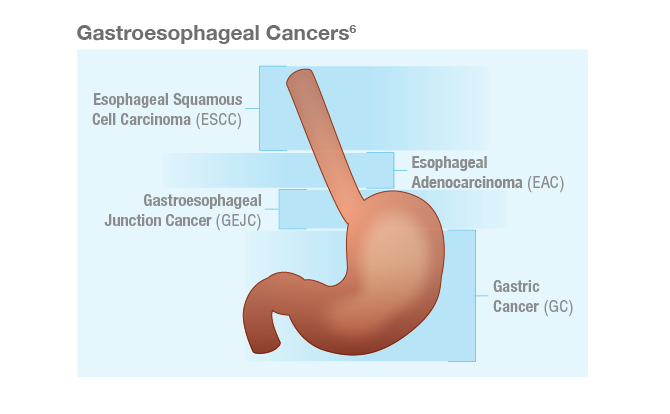
Introduction: Overview of gastroesophageal cancers

Gastroesophageal cancers consist of a group of heterogeneous tumors, including gastric cancer (GC), gastroesophageal junction cancer (GEJC), and esophageal cancer (EC).1 The majority of GC and GEJC are adenocarcinomas, while EC is categorized into 2 main histological subtypes: esophageal adenocarcinoma (EAC) and esophageal squamous cell carcinoma (ESCC).2,3 EAC is the predominant histology in the United States, contributing to ~62% of all EC cases.3,4 EAC incidence rates have been increasing over the past 5 decades in Western countries.4 Recent trends in the United States also suggest increasing incidence rates of GC overall in young adults (<50 years old).5

Gastric and esophageal cancers can be aggressive diseases with 5-year relative survival rates of <6% in the metastatic setting in the United States.7,8 Worldwide, GC and EC represent the fourth and sixth most common causes of cancer-related deaths, respectively.5

Approximately 15%–20% of gastroesophageal adenocarcinomas overexpress human epidermal growth factor receptor 2 (HER2)9. In this article, we will focus on HER2-negative gastroesophageal adenocarcinomas. Historically, chemotherapy has been the standard of care for the 1L treatment in this setting.10 In 2021,chemoimmunotherapy combinations were approved for appropriate patients with certain types of gastroesophageal cancers.11,12

OPDIVO + chemotherapy in 1L metastatic GC/GEJC/EAC

Currently, OPDIVO + fluoropyrimidine- and platinum-containing chemotherapy (chemo) is the only 1L chemoimmunotherapy regimen approved in metastatic non-HER2+ GC, GEJC, and EAC regardless of PD-L1 (programmed death ligand 1) status.11,13,14 The combination was approved based on the results of Checkmate 649, a global phase 3 study in 1L metastatic GC/GEJC/EAC patients with ECOG performance status 0-1.11,13 Key exclusion criteria included known HER2+ status and untreated CNS metastases.11 The study recruited all eligible patients regardless of PD-L1 expression.11,13

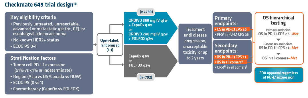
Checkmate 649 enrolled 1581 patients randomized 1:1 to OPDIVO + chemo (n=789) or chemo alone (n=792). A total of 473 patients in the OPDIVO + chemo arm and 482 patients in the chemo arm had tumors that expressed PD-L1 combined positive score (CPS) ≥5. The dual primary endpoints were overall survival (OS) and progression-free survival (PFS) in PD-L1 CPS ≥5. Key secondary endpoints included OS in PD-L1 CPS ≥1, OS in all randomized patients, and objective response rate (ORR) in all randomized patients. Checkmate 649 evaluated OPDIVO (10 mg/mL) injection for intravenous (IV) use (q2w or q3w) in combination with physician’s choice of either fluorouracil + oxaliplatin + leucovorin (mFOLFOX6) given q2w or capecitabine + oxaliplatin (CapeOx) given q3w. OPDIVO dosing was aligned with chemotherapy schedule. Treatment continued until disease progression, unacceptable toxicity, or up to 2 years. Baseline characteristics were consistent between all randomized and PD-L1 CPS ≥5 patients.13

There are Warnings and Precautions associated with OPDIVO to keep in mind. These include severe and fatal immune-mediated adverse reactions, infusion-related reactions, complications of allogeneic hematopoietic stem cell transplantation (HSCT); embryo-fetal toxicity, and increased mortality in patients with multiple myeloma when OPDIVO is added to a thalidomide analogue and dexamethasone, which is not recommended outside of controlled clinical trials.11 Additional information related to Warnings and Precautions can be found in the Important Safety Information below.

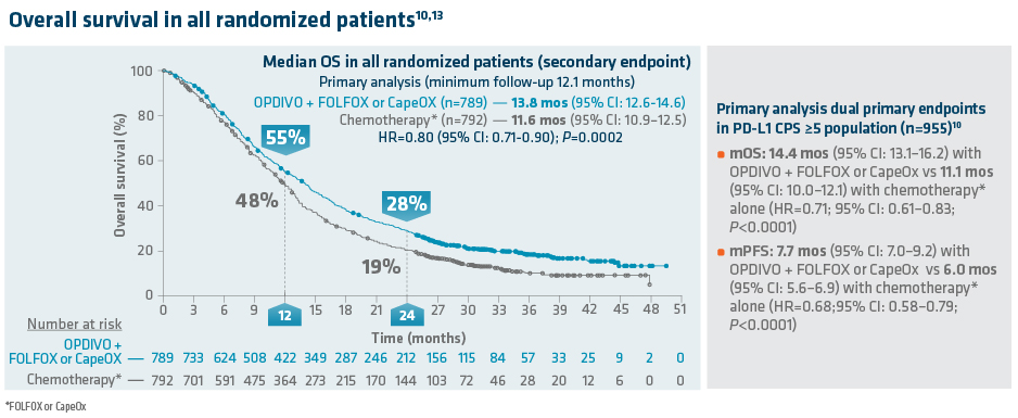
In the primary analysis (minimum[min] follow-up of 12.1 months[mos]), OPDIVO + chemo demonstrated superior OS in all randomized, PD-L1 CPS ≥1, and PD-L1 CPS ≥5 patients as compared to chemotherapy alone. In all randomized patients, mOS was 13.8 mos (95% confidence interval [CI]: 12.6–14.6) with OPDIVO + chemo vs 11.6 mos (95% CI: 10.9–12.5) with chemo (HR=0.80;95% CI: 0.71–0.90; P=0.0002). In PD-L1 CPS≥1 (n=1296), mOS was 14.0 mos (95% CI: 12.6–15.0) with OPDIVO + chemo vs 11.3 mos (95% CI: 10.6–12.3) with chemo (HR=0.77; 95% CI: 0.68–0.88; P<0.0001). In PD-L1 CPS≥5 (n=955), mOS was 14.4 mos (95% CI: 13.1–16.2) with OPDIVO + chemo vs 11.1 mos (95% CI: 10.0–12.1) with chemo (HR=0.71; 95% CI: 0.61–0.83; P<0.0001).11 The dual primary endpoint, mPFS in CPS ≥5 patients, was 7.7 mos (95% CI: 7.0–9.2) with OPDIVO + chemo vs 6.0 mos (95% CI: 5.6–6.9) with chemo (HR=0.68; 95% CI: 0.58–0.79; P<0.0001).

*FOLFOX or CapeOx.11†Assessed using blinded independent central review (BICR).11 ‡Based on confirmed response.11§Secondary endpoint.13

Exploratory OS analyses were reported for the primary (min follow-up 12.1 months) and follow-up (min follow-up 24 months) analysis. The 12-month OS rate in all randomized patients was 55% with OPDIVO + chemo vs 48% with chemo.13 The follow-up analysis at 24.0 months reported a mOS of 13.8 mos (95% CI: 12.4–14.5) with OPDIVO + chemo vs 11.6 mos (95% CI: 10.9–12.5) with chemo in all randomized patients (HR=0.79; (95% CI: 0.71–0.88) and 14.4 mos (95% CI: 13.1–16.2) with OPDIVO + chemo vs 11.1 mos with chemo (95% CI: 10.0–12.1)  in PD-L1 CPS ≥5 (HR=0.70; (95% CI: 0.60–0.81).14 The 24.0-month OS rate was 28% vs 19% for OPDIVO + chemo vs chemo, respectively, in all randomized patients.14

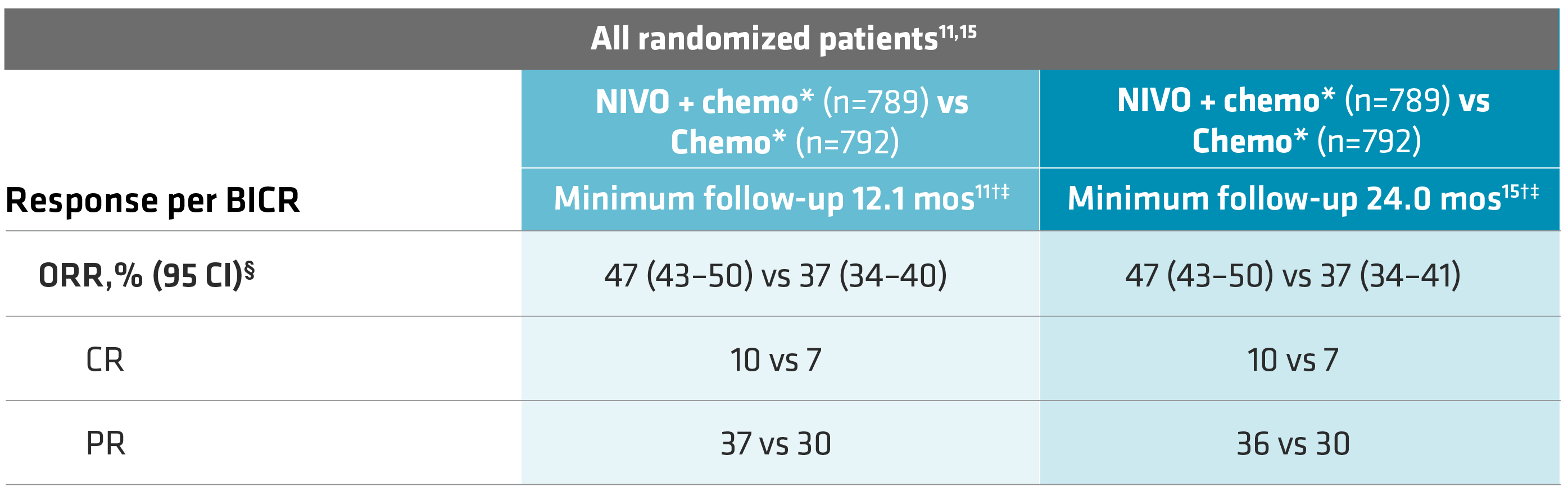
A secondary endpoint (min follow-up of 12.1 mos), ORR in all randomized patients, was 47% (95% CI: 43–50) with OPDIVO + chemo vs 37% (95% CI: 34–40) with chemo alone. Complete response (CR) rates were 10% vs 7% and partial response (PR) rates were 37% vs 30% for OPDIVO + chemo vs chemo, respectively.11

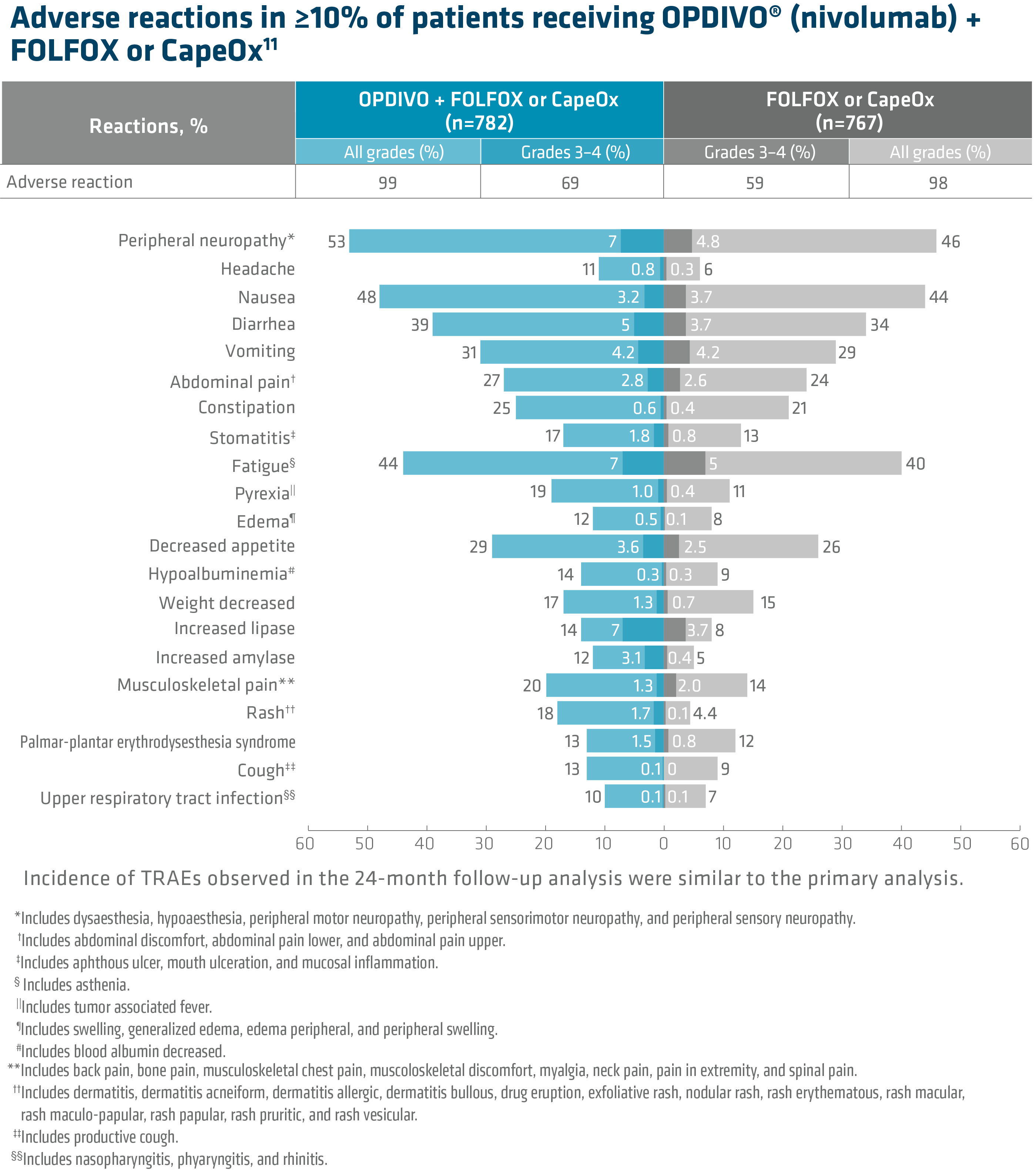
In Checkmate 649, the most common adverse reactions reported in ≥20% of patients treated with OPDIVO in combination with chemotherapy were peripheral neuropathy, nausea, fatigue, diarrhea, vomiting, decreased appetite, abdominal pain, constipation, and musculoskeletal pain. Of the ARs occurring in ≥10% of patients, those which were Grade 3–4 (OPDIVO + chemo vs chemo) were peripheral neuropathy (7% vs 4.8%), headache (0.8 vs 0.3%), nausea (3.2% vs 3.7%), diarrhea (5% vs 3.7%), vomiting (4.2% vs 4.2%), abdominal pain (2.8% vs 2.6%), constipation (0.6% vs 0.4%), stomatitis (1.8% vs 0.8%), fatigue (7% vs 5%), pyrexia (1% vs 0.4%), edema (0.5% vs 0.1%), decreased appetite (3.6% vs 2.5%), hypoalbuminemia (0.3% vs 0.3%), weight decreased (1.3% vs 0.7%), increased lipase (7% vs 3.7%), increased amylase (3.1% vs 0.4%), musculoskeletal pain (1.3% vs 2%), rash (1.7% vs 0.1%), palmar-plantar erythrodysesthesia syndrome (1.5% vs 0.8%), cough (0.1% vs 0%) and upper respiratory tract infection (0.1% vs 0.1%).

OPDIVO and/or chemotherapy were discontinued in 44% of patients and at least one dose was withheld in 76% of patients due to an adverse reaction. Serious adverse reactions occurred in 52% of patients treated with OPDIVO in combination with chemotherapy. The most frequent serious adverse reactions reported in ≥2% of patients treated with OPDIVO in combination with chemotherapy were vomiting (3.7%), pneumonia (3.6%), anemia (3.6%), pyrexia (2.8%), diarrhea (2.7%), febrile neutropenia (2.6%), and pneumonitis (2.4%). Fatal adverse reactions occurred in 16 (2.0%) patients who were treated with OPDIVO in combination with chemotherapy; these included pneumonitis (4 patients), febrile neutropenia (2 patients), stroke (2 patients), gastrointestinal toxicity, intestinal mucositis, septic shock, pneumonia, infection, gastrointestinal bleeding, mesenteric vessel thrombosis, and disseminated intravascular coagulation.11


Summary/conclusions

OPDIVO, in combination with fluoropyrimidine- and platinum-containing chemotherapy, is an approved treatment option for 1L metastatic non-HER2+ GC/GEJC/EAC regardless of PD-L1 status.11 This approval is based on the Checkmate 649 study, which at the primary analysis demonstrated superior OS with OPDIVO + chemotherapy versus chemotherapy in all randomized patients.11

1L=first line; chemo=chemotherapy; CI=confidence interval; CNS=central nervous system; ECOG=Eastern Cooperative Oncology Group; GEJC=gastroesophageal junction cancer; HR=hazard ratio; mo=month; mOS=median OS; q2w=every two weeks; q4w=every four weeks.

Indication

OPDIVO, in combination with fluoropyrimidine- and platinum-containing chemotherapy, is indicated for the treatment of patients with advanced or metastatic gastric cancer, gastroesophageal junction cancer, and esophageal adenocarcinoma.11

Important Safety Information

Severe and Fatal Immune-Mediated Adverse Reactions

• Immune-mediated adverse reactions listed herein may not include all possible severe and fatal immune-mediated adverse reactions.
• Immune-mediated adverse reactions, which may be severe or fatal, can occur in any organ system or tissue. While immune-mediated adverse reactions usually manifest during treatment, they can also occur after discontinuation of OPDIVO. Early identification and management are essential to ensure safe use of OPDIVO. Monitor for signs and symptoms that may be clinical manifestations of underlying immune-mediated adverse reactions. Evaluate clinical chemistries including liver enzymes, creatinine, and thyroid function at baseline and periodically during treatment with OPDIVO. In cases of suspected immune-mediated adverse reactions, initiate appropriate workup to exclude alternative etiologies, including infection. Institute medical management promptly, including specialty consultation as appropriate.
• Withhold or permanently discontinue OPDIVO depending on severity (please see section 2 Dosage and Administration in the accompanying Full Prescribing Information). In general, if OPDIVO interruption or discontinuation is required, administer systemic corticosteroid therapy (1 to 2 mg/kg/day prednisone or equivalent) until improvement to Grade 1 or less. Upon improvement to Grade 1 or less, initiate corticosteroid taper and continue to taper over at least 1 month. Consider administration of other systemic immunosuppressants in patients whose immune-mediated adverse reactions are not controlled with corticosteroid therapy. Toxicity management guidelines for adverse reactions that do not necessarily require systemic steroids (e.g., endocrinopathies and dermatologic reactions) are discussed below.

Immune-Mediated Pneumonitis
• OPDIVO can cause immune-mediated pneumonitis. The incidence of pneumonitis is higher in patients who have received prior thoracic radiation. In patients receiving OPDIVO monotherapy, immune-mediated pneumonitis occurred in 3.1% (61/1994) of patients, including Grade 4 (<0.1%), Grade 3 (0.9%), and Grade 2 (2.1%).

Immune-Mediated Colitis
• OPDIVO can cause immune-mediated colitis. A common symptom included in the definition of colitis was diarrhea. Cytomegalovirus (CMV) infection/reactivation has been reported in patients with corticosteroid-refractory immune-mediated colitis. In cases of corticosteroid-refractory colitis, consider repeating infectious workup to exclude alternative etiologies. In patients receiving OPDIVO monotherapy, immune-mediated colitis occurred in 2.9% (58/1994) of patients, including Grade 3 (1.7%) and Grade 2 (1%).

Immune-Mediated Hepatitis and Hepatoxicity
• OPDIVO can cause immune-mediated hepatitis. In patients receiving OPDIVO monotherapy, immune-mediated hepatitis occurred in 1.8% (35/1994) of patients, including Grade 4 (0.2%), Grade 3 (1.3%), and Grade 2 (0.4%).

Immune-Mediated Endocrinopathies
• OPDIVO can cause primary or secondary adrenal insufficiency, immune-mediated hypophysitis, immune-mediated thyroid disorders, and Type 1 diabetes mellitus, which can present with diabetic ketoacidosis. Withhold OPDIVO depending on severity (please see section 2 Dosage and Administration in the accompanying Full Prescribing Information). For Grade 2 or higher adrenal insufficiency, initiate symptomatic treatment, including hormone replacement as clinically indicated. Hypophysitis can present with acute symptoms associated with mass effect such as headache, photophobia, or visual field defects. Hypophysitis can cause hypopituitarism; initiate hormone replacement as clinically indicated. Thyroiditis can present with or without endocrinopathy. Hypothyroidism can follow hyperthyroidism; initiate hormone replacement or medical management as clinically indicated. Monitor patients for hyperglycemia or other signs and symptoms of diabetes; initiate treatment with insulin as clinically indicated.
• In patients receiving OPDIVO monotherapy, adrenal insufficiency occurred in 1% (20/1994), including Grade 3 (0.4%) and Grade 2 (0.6%).
• In patients receiving OPDIVO monotherapy, hypophysitis occurred in 0.6% (12/1994) of patients, including Grade 3 (0.2%) and Grade 2 (0.3%).
• In patients receiving OPDIVO monotherapy, thyroiditis occurred in 0.6% (12/1994) of patients, including Grade 2 (0.2%).
• In patients receiving OPDIVO monotherapy, hyperthyroidism occurred in 2.7% (54/1994) of patients, including Grade 3 (<0.1%) and Grade 2 (1.2%).
• In patients receiving OPDIVO monotherapy, hypothyroidism occurred in 8% (163/1994) of patients, including Grade 3 (0.2%) and Grade 2 (4.8%).
• In patients receiving OPDIVO monotherapy, diabetes occurred in 0.9% (17/1994) of patients, including Grade 3 (0.4%) and Grade 2 (0.3%), and 2 cases of diabetic ketoacidosis.

Immune-Mediated Nephritis with Renal Dysfunction
• OPDIVO can cause immune-mediated nephritis. In patients receiving OPDIVO® monotherapy, immune-mediated nephritis and renal dysfunction occurred in 1.2% (23/1994) of patients, including Grade 4 (<0.1%), Grade 3 (0.5%), and Grade 2 (0.6%).

Immune-Mediated Dermatologic Adverse Reactions
• OPDIVO can cause immune-mediated rash or dermatitis. Exfoliative dermatitis, including Stevens-Johnson syndrome (SJS), toxic epidermal necrolysis (TEN), and drug rash with eosinophilia and systemic symptoms (DRESS) has occurred with PD-1/PD-L1 blocking antibodies. Topical emollients and/or topical corticosteroids may be adequate to treat mild to moderate nonexfoliative rashes.
• Withhold or permanently discontinue OPDIVO depending on severity (please see section 2 Dosage and Administration in the accompanying Full Prescribing Information).
• In patients receiving OPDIVO monotherapy, immune-mediated rash occurred in 9% (171/1994) of patients, including Grade 3 (1.1%) and Grade 2 (2.2%).

Other Immune-Mediated Adverse Reactions
• The following clinically significant immune-mediated adverse reactions occurred at an incidence of <1% (unless otherwise noted) in patients who received OPDIVO monotherapy or were reported with the use of other PD-1/PD-L1 blocking antibodies. Severe or fatal cases have been reported for some of these adverse reactions: cardiac/vascular: myocarditis, pericarditis, vasculitis; nervous system: meningitis, encephalitis, myelitis and demyelination, myasthenic syndrome/myasthenia gravis (including exacerbation), Guillain-Barré syndrome, nerve paresis, autoimmune neuropathy; ocular: uveitis, iritis, and other ocular inflammatory toxicities can occur; gastrointestinal: pancreatitis to include increases in serum amylase and lipase levels, gastritis, duodenitis; musculoskeletal and connective tissue: myositis/polymyositis, rhabdomyolysis, and associated sequelae including renal failure, arthritis, polymyalgia rheumatica; endocrine: hypoparathyroidism; other (hematologic/immune): hemolytic anemia, aplastic anemia, hemophagocytic lymphohistiocytosis (HLH), systemic inflammatory response syndrome, histiocytic necrotizing lymphadenitis (Kikuchi lymphadenitis), sarcoidosis, immune thrombocytopenic purpura, solid organ transplant rejection.
• Some ocular IMAR cases can be associated with retinal detachment. Various grades of visual impairment, including blindness, can occur. If uveitis occurs in combination with other immune-mediated adverse reactions, consider a Vogt-Koyanagi-Harada–like syndrome, which has been observed in patients receiving OPDIVO, as this may require treatment with systemic corticosteroids to reduce the risk of permanent vision loss.

Infusion-Related Reactions
• OPDIVO can cause severe infusion-related reactions. Discontinue OPDIVO in patients with severe (Grade 3) or life-threatening (Grade 4) infusion-related reactions. Interrupt or slow the rate of infusion in patients with mild (Grade 1) or moderate (Grade 2) infusion-related reactions. In patients receiving OPDIVO monotherapy as a 60-minute infusion, infusion-related reactions occurred in 6.4% (127/1994) of patients. In a separate trial in which patients received OPDIVO monotherapy as a 60-minute infusion or a 30-minute infusion, infusion-related reactions occurred in 2.2% (8/368) and 2.7% (10/369) of patients, respectively. Additionally, 0.5% (2/368) and 1.4% (5/369) of patients, respectively, experienced adverse reactions within 48 hours of infusion that led to dose delay, permanent discontinuation or withholding of OPDIVO.
Complications of Allogeneic Hematopoietic Stem Cell Transplantation
• Fatal and other serious complications can occur in patients who receive allogeneic hematopoietic stem cell transplantation (HSCT) before or after being treated with OPDIVO. Transplant-related complications include hyperacute graft-versus-host-disease (GVHD), acute GVHD, chronic GVHD, hepatic veno-occlusive disease (VOD) after reduced intensity conditioning, and steroid-requiring febrile syndrome (without an identified infectious cause). These complications may occur despite intervening therapy between OPDIVO and allogeneic HSCT.
• Follow patients closely for evidence of transplant-related complications and intervene promptly. Consider the benefit versus risks of treatment with OPDIVO prior to or after an allogeneic HSCT.
Embryo-Fetal Toxicity
• Based on its mechanism of action and findings from animal studies, OPDIVO can cause fetal harm when administered to a pregnant woman. Advise pregnant women of the potential risk to a fetus. Advise females of reproductive potential to use effective contraception during treatment with OPDIVO and for at least 5 months after the last dose.
Increased Mortality in Patients with Multiple Myeloma when OPDIVO® is Added to a Thalidomide Analogue and Dexamethasone
• In randomized clinical trials in patients with multiple myeloma, the addition of OPDIVO to a thalidomide analogue plus dexamethasone resulted in increased mortality. Treatment of patients with multiple myeloma with a PD-1 or PD-L1 blocking antibody in combination with a thalidomide analogue plus dexamethasone is not recommended outside of controlled clinical trials.
Lactation
• There are no data on the presence of OPDIVO in human milk, the effects on the breastfed child, or the effects on milk production. Because of the potential for serious adverse reactions in breastfed children, advise women not to breastfeed during treatment and for 5 months after the last dose.
Serious Adverse Reactions
• In Checkmate 649, serious adverse reactions occurred in 52% of patients treated with OPDIVO in combination with chemotherapy (n=782). The most frequent serious adverse reactions reported in ≥2% of patients treated with OPDIVO in combination with chemotherapy were vomiting (3.7%), pneumonia (3.6%), anemia (3.6%), pyrexia (2.8%), diarrhea (2.7%), febrile neutropenia (2.6%), and pneumonitis (2.4%). Fatal adverse reactions occurred in 16 (2.0%) patients who were treated with OPDIVO in combination with chemotherapy; these included pneumonitis (4 patients), febrile neutropenia (2 patients), stroke (2 patients), gastrointestinal toxicity, intestinal mucositis, septic shock, pneumonia, infection, gastrointestinal bleeding, mesenteric vessel thrombosis, and disseminated intravascular coagulation.
Common Adverse Reactions
• In Checkmate 649, the most common adverse reactions (≥20%) in patients treated with OPDIVO in combination with chemotherapy (n=782) were peripheral neuropathy (53%), nausea (48%), fatigue (44%), diarrhea (39%), vomiting (31%), decreased appetite (29%), abdominal pain (27%), constipation (25%), and musculoskeletal pain (20%).

Please see US Full Prescribing Information for OPDIVO.

References:

1. Paydary K, Reizine N, Catenacci DVT. Immune-checkpoint inhibition in the treatment of gastro-esophageal cancer: a closer look at the emerging evidence. Cancers (Basel). 2021;13(23):5929.
2. National Cancer Institute. Gastric cancer treatment (PDQ®)–health professional version. National Cancer Institute website. Updated April 22, 2021.Accessed December 3, 2021.
http://cancer.gov/types/stomach/hp/stomach-treatment-pdq.
3. Chen Z, Ren Y, Du XL, et al. Incidence and survival differences in esophageal cancer among ethnic groups in the United States. Oncotarget. 2017;8(29):47037-47051.
4. He H, Chen N, Hou Y, et al. Trends in the incidence and survival of patients with esophageal cancer: a SEER database analysis. Thorac Cancer. 2020;11(5):1121-1128.
5. Sung H, Ferlay J, Siegel RL, et al. Global cancer statistics 2020: GLOBOCAN estimates of incidence and mortality worldwide for 36 cancers in 185 countries.CA Cancer J Clin. 2021;71(3):209-249.
6. Arnold M, Ferlay J, van Berge Henegouwen MI, Soerjomataram I. Global burden of oesophageal and gastric cancer by histology and subsite in 2018. Gut. 2020;69(9):1564-1571.
7. National Cancer Institute Surveillance, Epidemiology, and End Results Program. Cancer stat facts: stomach cancer. National Cancer Institute website. Accessed December 3, 2021.
http://seer.cancer.gov/statfacts/html/stomach.html.
8. National Cancer Institute Surveillance, Epidemiology, and End Results Program. Cancer stat facts: esophageal cancer. National Cancer Institute website. Accessed December 3, 2021.
http://seer.cancer.gov/statfacts/html/esoph.html.
9. Grieb BC, Agarwal R. HER2-Directed Therapy in Advanced Gastric and Gastroesophageal Adenocarcinoma: Triumphs and Troubles. Curr Treat Options Oncol. 2021;22(10):88.
10. ShankaranV, Xiao, H, Bertwistle D, et al. A comparison of real-world treatment patterns and clinical outcomes in patients receiving first-line therapy for unresectable advanced gastric or gastroesophageal junction cancer versus esophageal adenocarcinomas. Adv Ther. 2021;38:
707-720.
11. OPDIVO® (nivolumab) [package insert]. Princeton, NJ: Bristol-Myers Squibb Company; 2021.
12. KEYTRUDA® (pembrolizumab) [package insert]. Kenilworth, NJ: Merck & Co., Inc; 2021.
13. Janjigian YY, Shitara K, Moehler M, et al. First-line nivolumab plus chemotherapy versus chemotherapy alone for advanced gastric, gastroesophageal junction cancer/oesophageal adenocarcinoma (CheckMate 649): a randomised, open-label, phase 3 trial. Lancet. 2021;398(10294):27-40.
14. Janjigian YY, Ajani JA, Moehler M, et al. Nivolumab plus chemotherapy or ipilimumab vs chemotherapy as first-line treatment for advanced gastric cancer/gastroesophageal junction cancer/ esophageal adenocarcinoma: CheckMate 649 study. Presentation at ESMO 2021. Abstract LBA7.
15. Data on file. BMS-REF-NIVO-0120. Princeton, NJ: Bristol-Myers Squibb Company; 2021.

[email protected]
Please click here to unsubscribe. Opting out of the subscription service emails will not opt you out of messages from Bristol-Myers Squibb Company.
©2022 Bristol-Myers Squibb Company. All rights reserved. OPDIVO® and the related logos are registered trademarks of Bristol-Myers Squibb Company.
1506-US-2200006 03/22

TUKYSA® in Pretreated HER2-positive Metastatic Breast Cancer With and Without Brain Metastases: Final Overall Survival Analysis

SUMMARY: Breast cancer is the most common cancer among women in the US and about 1 in 8 women (12%) will develop invasive breast cancer during their lifetime. Approximately 290,560 new cases of breast cancer will be diagnosed in 2022 and about 43,780 individuals will die of the disease, largely due to metastatic recurrence.

The HER or erbB family of receptors consist of HER1, HER2, HER3 and HER4. Approximately 15-20% of invasive breast cancers overexpress HER2/neu oncogene, which is a negative predictor of outcomes without systemic therapy. Patients with HER2-positive metastatic breast cancer are often treated with anti-HER2 targeted therapy along with chemotherapy, irrespective of hormone receptor status, and this has resulted in significantly improved treatment outcomes. HER2-targeted therapies include HERCEPTIN® (Trastuzumab), TYKERB® (Lapatinib), PERJETA® (Pertuzumab), KADCYLA® (ado-Trastuzumab emtansine), ENHERTU® (Trastuzumab deruxtecan) and MARGENZA® (Margetuximab). Dual HER2 blockade with HERCEPTIN® and PERJETA®, given along with chemotherapy (with or without endocrine therapy), as first line treatment, in HER2-positive metastatic breast cancer patients, was shown to significantly improve Progression Free Survival (PFS) as well as Overall Survival (OS). The superior benefit with dual HER2 blockade has been attributed to differing mechanisms of action and synergistic interaction between HER2 targeted therapies. Patients progressing on Dual HER2 blockade often receive KADCYLA® which results in an Objective Response Rate (ORR) of 44% and a median PFS of 9.6 months, when administered after HERCEPTIN® and a taxane. There is however no standard treatment option for this patient population following progression on KADCYLA®.

It is estimated that close to 50% of patients with HER2-positive metastatic breast cancer develop brain metastases. Systemic HER2-targeted agents, including Tyrosine Kinase Inhibitors, as well as chemotherapy have limited antitumor activity in the brain. This is therefore an area of high unmet need. Local therapeutic interventions for brain metastases include neurosurgical resection and Stereotactic or Whole-Brain Radiation Therapy.

TUKYSA® (Tucatinib) is an oral Tyrosine Kinase Inhibitor that is highly selective for the kinase domain of HER2 with minimal inhibition of Epidermal Growth Factor Receptor. In a Phase 1b dose-escalation trial, TUKYSA® in combination with HERCEPTIN® and XELODA® (Capecitabine) showed encouraging antitumor activity in patients with HER2-positive metastatic breast cancer, including those with brain metastases.

HER2CLIMB is an international, randomized, double-blind, placebo-controlled trial in which the combination of TUKYSA® plus HERCEPTIN® and XELODA® was compared with placebo plus HERCEPTIN® and XELODA®. A total of 612 patients with unresectable locally advanced or metastatic HER2-positive breast cancer, who were previously treated with HERCEPTIN®, PERJETA® (Pertuzumab) and KADCYLA® (ado-Trastuzumab emtansine) were enrolled. Patients were randomly assigned in a 2:1 ratio to receive either TUKYSA® 300 mg orally twice daily throughout the treatment period (N=410) or placebo orally twice daily (N=201), in combination with HERCEPTIN® 6 mg/kg IV once every 21 days, following an initial loading dose of 8 mg/kg, and XELODA® 1000 mg/m2 orally twice daily on days 1 to 14 of each 21-day cycle. Stratification factors included presence or absence of brain metastases, ECOG Performance Status and geographic region. The median patient age was 54 years and patient demographic as well as disease characteristics at baseline were well balanced between the two treatment groups. In the total treatment population, 47.5% had brain metastases at baseline, 48.3% in the TUKYSA® combination group and 46% in the placebo combination group. The Primary endpoint was Progression Free Survival (PFS). Secondary end points included Overall Survival (OS), PFS among patients with brain metastases, confirmed Objective Response Rate (ORR), and safety.

In the primary analysis, at a median follow-up of 14 months, TUKYSA® added to HERCEPTIN® and XELODA®, significantly improved Overall Survival (OS) and Progression Free Survival (PFS) in patients with HER2-positive metastatic breast cancer. After the primary analysis, the protocol was amended to allow unblinding and cross-over from the placebo combination to the TUKYSA® combination. Protocol prespecified descriptive analyses of OS, PFS and safety were carried out at about 2 years from the last patient randomized. The researchers in this publication reported the final efficacy and safety outcomes after an additional 15.6 months follow up (total follow up of 29.6 months) in patients from the HER2CLIMB trial.

At a median follow up of 29.6 months, the median duration of OS was 24.7 months for the TUKYSA® combination group versus 19.2 months in the placebo combination group (HR for death=0.73; P=0.004). The estimated OS rate at 2 years was 51% in the TUKYSA® combination group and 40% in the placebo combination group. The OS benefit with the TUKYSA® combination was noted across all prespecified subgroups in the overall study population and was consistent with the primary analysis. The median duration of PFS was 7.6 months for the TUKYSA® combination group versus 4.9 months for the placebo combination group (HR for progression or death=0.57; P<0.00001), and PFS at 1 year was 29% and 14%, respectively.

Systemic treatment with TUKYSA® in combination with HERCEPTIN® and XELODA® provided consistent clinical benefit to patients with and without brain metastases. TUKYSA® combination doubled the intracranial Objective Response Rate, reduced the risk of intracranial progression or death by two-thirds in all patients with brain metastases. In this study population, the estimated 1-year intracranial PFS was 40% in the TUKYSA® group and 0% in the control group. In patients with untreated or treated and progressing (active) brain metastases, the estimated 1-year intracranial PFS was 35% in the TUKYSA® group, 0% in the control group, and in patients with treated (stable) brain metastases, was 53% in the TUKYSA® group and 0% in the control group. The TUKYSA® combination was well tolerated with a low rate of discontinuation due to toxicities. Common adverse events in the TUKYSA® group included diarrhea, Palmar-Plantar Erythrodysesthesia syndrome, nausea, vomiting and fatigue. Diarrhea and abnormal liver function tests were more common in the TUKYSA® group than in the control group.

It was concluded that with additional follow up, TUKYSA® in combination with HERCEPTIN® and XELODA® provided a clinically meaningful survival benefit, including those with brain metastases, supporting the use of this combination in patients with previously treated HER2-positive metastatic breast cancer, after progression on two HER2-targeted therapies.

Tucatinib versus placebo added to trastuzumab and capecitabine for patients with pretreated HER2+ metastatic breast cancer with and without brain metastases (HER2CLIMB): final overall survival analysis. Curigliano G, Mueller V, Borges V, et al. Ann Oncol. 2022;33:321-329.

Early-Stage Multi-Cancer Detection Using an Extracellular Vesicle Protein-Based Blood Test

SUMMARY: The American Cancer Society’s estimates that in 2022, about 62,210 people will be diagnosed with pancreatic cancer and 49,830 people will die of the disease, 19,880 women will receive a new diagnosis of ovarian cancer and about 12,810 women will die of the disease, and about 81,800 new cases of bladder cancer will be diagnosed in 2022 and about 17,100 patients will die of the disease. These three cancer types are estimated to account for approximately 80,000 deaths in the US in 2022. Detecting cancer at early stages can significantly increase survival rates and outcomes.

Several multi-cancer early detection tests are being developed that involve blood-based circulating cell-free tumor DNA (cfDNA) in the plasma, to track hundreds of patient-specific mutations, to detect Minimal Residual Disease (MRD) , as well as detection of abnormal methylation patterns, followed by machine learning approaches, to differentiate between cancer and non-cancer, for detecting clinically significant, late-stage (III and IV) cancers. Early detection of cancer however is the key to improving survival. This is particularly relevant for certain cancer types. Pancreatic Ductal AdenoCarcinoma (PDAC) is one of the deadliest cancers, and a leading cause of all cancer-related deaths in the United States, and is typically detected when the disease is advanced. However, when detected at Stage I, survival rates can be as high as 80%. Ovarian cancer is often detected when the disease is advanced and the 5-year survival rates are less than 30%, but can be as high as 93% when detected early. The same holds true for metastatic bladder cancer, with 5-year survival rates of only 6%, whereas while detected when the tumor is still localized to the bladder wall inner layer results in a 5-year survival rate of 96%. Even though serum CA19-9 is intended as an aid in the management of patients with confirmed pancreatic cancer for serial monitoring of their response to therapy and disease progression, it is not recommended by the FDA for screening, as it may be elevated in several benign conditions. Similarly, serum CA-125 is FDA approved for use in monitoring patients with ovarian cancer for disease persistence and recurrence, but is not recommended to screen for ovarian cancer. Currently, there are few general screening strategies to detect asymptomatic, early-stage PDAC, ovarian, or bladder cancer and there is therefore a significant unmet need in this patient group.

Exosomes are 30-150 nm-sized Extracellular Vesicles (EVs) secreted by multiple different cell types and ejected by tumors into the bloodstream. They mediate intercellular signaling by transferring mRNAs and microRNAs between distant cells and tissues and therefore carry functional protein biomarkers representing the tumor proteome. Exosomes represent one potential approach for more sensitive detection of cancer-related biomarkers from blood.

The researchers in this study used an Alternating Current Electrokinetic (ACE)-based platform (Verita™ System) to efficiently isolate EVs from soluble contaminants such as cells, small proteins, or other vesicles from patient samples, and then measured the concentrations of associated protein biomarkers (“EV proteins”) present in the purified EV samples from our case-control study subjects. The researchers chose this platform over the current gold standard ultracentrifugation method, which the authors felt was inefficient and not suitable for point-of-care applications. Artificial Intelligence machine-learning algorithm developed by the researchers, enabled detection of early-stage pancreatic, ovarian, and bladder cancers.

In this case-control pilot study, 139 pathologically confirmed Stage I and II cancer cases representing pancreatic, ovarian, or bladder patients were compared with 184 control subjects, using the Verita™ System. The Extracellular Vesicles (EVs) isolated using this technology, were consistent with the presence of Exosomes, in accordance with the International Society for Extracellular Vesicles (ISEV) 2018 guidelines. The researchers selected a panel of 13 Extracellular Vesicle (EV) proteins along with age, a known cofactor in cancer. In order to simulate a real-world screening scenario, all cancer cases were treatment-naïve and to ensure that these were early-stage patients, the histopathologic staging was confirmed using the American Joint Commission on Cancer (AJCC) guidelines. The median age of the cancer cases was 60 years and 63.3% of the overall cancer cases were Stage I, with the remaining 36.7% at Stage II. The median age of the control group was 57 years and had no known history of cancer, autoimmune diseases, neurodegenerative disorders or diabetes mellitus.

When the overall cancer case cohort was compared with the control individuals using the EV protein biomarker test, the average sensitivity was 71.2%, at a specificity of 99.5%. When considered across all the three cancers studied, EV protein biomarker test using this technology demonstrated similar sensitivities of 70.5% and 72.5% for Stage I and II patients, respectively. This new technology detected 95.5% of Stage I pancreatic cancers, 73.1% of pathologic Stage IA lethally aggressive serous ovarian adenocarcinomas and 43.8% in bladder cancer, demonstrating the potential value of this platform for detection of early stage cancers. The lower sensitivity for detecting early stage bladder cancer may be due to high molecular and histologic heterogeneity of bladder tumors.

It was concluded from this study that blood-based EV protein detection test has potential clinical value for early cancer detection and the use of Verita™ platform resulted in the accurate detection of early stage pancreatic, ovarian, or bladder cancer. The authors added that mortality from pancreatic cancer which will soon become the second leading cause of cancer mortality in the U.S., can be greatly reduced if this study results are validated.

Early-stage multi-cancer detection using an extracellular vesicle protein-based blood test. Hinestrosa, J.P., Kurzrock, R., Lewis, J.M. et al. Commun Med 2, 29 (2022). https://doi.org/10.1038/s43856-022-00088-6.

FDA Approves Neoadjuvant OPDIVO® and Chemotherapy Combination for Early Stage Non Small Cell Lung Cancer

SUMMARY: The FDA on March 4, 2022, approved OPDIVO® (Nivolumab) with platinum-doublet chemotherapy for adult patients with resectable Non Small Cell Lung Cancer (NSCLC) in the neoadjuvant setting. This represents the first FDA approval for neoadjuvant therapy for early stage NSCLC. The American Cancer Society estimates that for 2022, about 236,740 new cases of lung cancer will be diagnosed and 135,360 patients will die of the disease. Lung cancer is the leading cause of cancer-related mortality in the United States. Non-Small Cell Lung Cancer (NSCLC) accounts for approximately 85% of all lung cancers. Of the three main subtypes of NSCLC, 30% are Squamous Cell Carcinomas (SCC), 40% are Adenocarcinomas and 10% are Large Cell Carcinomas. With changes in the cigarette composition and decline in tobacco consumption over the past several decades, Adenocarcinoma now is the most frequent histologic subtype of lung cancer.

Surgical resection with a curative intent is the primary treatment for approximately 30% of patients with NSCLC who present with early Stage (I–IIIA) disease, unless medically unfit. These numbers are likely to increase with the implementation of Lung Cancer screening programs. These patients are often treated with platinum-based adjuvant chemotherapy/immunotherapy following surgical resection, to decrease the risk of recurrence. Nonetheless, 45-75% of these patients develop recurrent disease. There is therefore an unmet need for this patient population.

CHECKMATE-816 is an open-label, multicenter, randomized Phase III study which evaluated OPDIVO® plus chemotherapy versus chemotherapy alone as neoadjuvant treatment in patients with resectable Stage IB to IIIA NSCLC. In this trial, 358 patients with clinical Stage IB to Stage IIIA resectable NSCLC, with an ECOG Performance Status of 0 to 1 and no known sensitizing EGFR mutations or ALK alterations, were randomly assigned 1:1 to receive OPDIVO® at a dose of 360 mg IV along with platinum-doublet chemotherapy every 3 weeks for 3 doses (N=179) or chemotherapy alone on the same schedule (N=179). Patients then underwent radiologic staging and surgical resection within 6 weeks of neoadjuvant therapy. They then had the option of adjuvant therapy with or without radiation therapy, and were followed up. Both treatment groups were well balanced with regards to age, sex, histology and smoking status. About two-thirds of the patients had Stage IIIA disease. The median patient age was 65 years and patients were stratified by cancer stage, gender and PD-L1 status (1% or higher versus less than 1%). Tumor Mutational Burden results were available for 50% of patients. The Primary end points of this study were pathologic Complete Response (pCR), defined as the absence of viable tumor cells in lung and lymph nodes, and Event Free Survival (EFS). Secondary endpoints include major pathological response and Overall Survival. Key exploratory endpoints included feasibility of surgery and surgery-related adverse events.

The pCR rate was 24% in the OPDIVO® plus chemotherapy group and 2.2% in the chemotherapy alone group. The pCR improvement was noted with the OPDIVO® plus chemotherapy combination regardless of disease stage and irrespective of radiologic downstaging. Overall, 83% of patients assigned to OPDIVO® plus chemotherapy and 78% of patient’s assigned to chemotherapy alone achieved R0 resection, with 10% versus 74% median residual viable tumor cells noted in the primary tumor bed respectively. Lung-sparing surgery (lobectomy) was performed in 77% of patients assigned to OPDIVO® plus chemotherapy versus 61% among those assigned to chemotherapy alone. The median EFS was 31.6 months in the OPDIVO® plus chemotherapy group and 20.8 months for those receiving chemotherapy alone (HR=0.63; P=0.0052).

The authors concluded that CheckMate 816 is the first Phase III trial to show a benefit for neoadjuvant immunotherapy plus platinum-doublet chemotherapy in earlier stage resectable NSCLC, with marked improvement in pathologic Complete Response rate, without any meaningful increase in toxicity or decrease in the feasibility of surgery. It is likely that the higher pathologic Complete Response rate may translate into higher cure rates, with longer follow up.

Surgical outcomes from the phase 3 CheckMate 816 trial: nivolumab (NIVO) + platinum-doublet chemotherapy (chemo) vs chemo alone as neoadjuvant treatment for patients with resectable non-small cell lung cancer (NSCLC). Spicer J, Wang C, Tanaka F, et al. J Clin Oncol. 2021;39(suppl 15):8503. doi:10.1200/JCO.2021.39.15_suppl.8503

FDA Approves YESCARTA® for Second Line Treatment of Large B-cell Lymphoma

SUMMARY: The FDA on April 1, 2022, approved YESCARTA® (Axicabtagene ciloleucel) for adult patients with Large B-cell lymphoma (LBCL) that is refractory to first-line chemoimmunotherapy or relapses within 12 months of first-line chemoimmunotherapy.

What is (CAR) T-cell immunotherapy? Chimeric Antigen Receptor (CAR) T-cell therapy is a type of immunotherapy and consists of T cells collected from the patient’s blood in a leukapheresis procedure, and genetically engineered to produce special receptors on their surface called Chimeric Antigen Receptors (CAR). These reprogrammed cytotoxic T cells with the Chimeric Antigen Receptors on their surface are now able to recognize a specific antigen on tumor cells. These genetically engineered and reprogrammed CAR T-cells are grown in the lab and are then infused into the patient. These cells in turn proliferate in the patient’s body and the engineered receptor on the cell surface help recognize and kill cancer cells that expresses that specific antigen. It is a therefore a customized treatment created using patient’s own T cells to destroy cancer cells.Chimeric-Antigen-Receptor-T-Cell-Immunotherapy

YESCARTA® is a Chimeric Antigen Receptor (CAR) T cell immunotherapy and consists of autologous T cells that are genetically modified to produce a CAR protein, allowing the T cells to seek out and destroy cancer cells expressing the antigen CD19, which is found uniquely on B cells. Patients, following treatment with CAR T-cells, develop B-cell aplasia (absence of CD19 positive cells) due to B-cell destruction and may need immunoglobin replacement. Hence, B-cell aplasia can be a useful therapeutic marker, as continued B-cell aplasia has been seen in all patients who had sustained remission, following CAR T-cell therapy. Cytokine Release Syndrome (CRS), an inflammatory process is the most common and serious side effect of CAR T-cell therapy and is associated with marked elevation of Interleukin-6. Cytokine release is important for T-cell activation and can result in high fevers and myalgias. This is usually self limiting although if severe can be associated with hypotension and respiratory insufficiency. Tocilizumab (ACTEMRA®), an Interleukin-6 receptor blocking antibody produces a rapid improvement in symptoms. This is however not recommended unless the symptoms are severe and life threatening, as blunting the cytokine response can in turn negate T-cell proliferation. Elevated serum Ferritin and C-reactive protein levels are surrogate markers for severe Cytokine Release Syndrome. The CAR T-cells have been shown to also access sanctuary sites such as the central nervous system and eradicate cancer cells. CD19 antigen is expressed by majority of the B cell malignancies and therefore most studies using CAR T-cell therapy have focused on the treatment of advanced B-cell malignancies such as Chronic Lymphocytic Leukemia (CLL), Acute Lymphoblastic Leukemia (ALL) and Non Hodgkin lymphoma (NHL), such as Diffuse Large B-Cell Lymphoma (DLBCL).

Diffuse Large B-Cell Lymphoma (DLBCL) is the most common of the aggressive Non-Hodgkin lymphoma’s in the United States, and the incidence has steadily increased 3 to 4% each year. Outcomes for patients with relapsed/refractory disease, is poor, with an Objective response Rate (ORR) of 26%, Complete Response (CR) rate of 8% and a median Overall Survival (OS) of 6.6 months. There is therefore a significant unmet need in this patient group.

The present FDA approval was based on ZUMA-7, an international, randomized, open-label, multicenter Phase III trial, which compared the safety and efficacy of YESCARTA® with that of the current Standard of Care, as second-line treatment in patients with relapsed or refractory Large B-Cell Lymphoma (LBCL). In this study, 359 enrolled patients were randomized 1:1 to receive either a single infusion of YESCARTA® following Fludarabine and Cyclophosphamide lymphodepleting chemotherapy (N=180) or Standard of Care investigator-chosen second-line therapy, consisting of 2 or 3 cycles of chemoimmunotherapy followed by high-dose therapy and autologous Hematopoietic Stem Cell Transplantation (HSCT), in patients who attained Complete Remission or Partial Remission (N=179). Patients randomized to YESCARTA® underwent leukapheresis, and then, lymphodepleting chemotherapy with Fludarabine 30 mg/m2/day and Cyclophosphamide 500 mg/m2/day for 3 days, followed by a single infusion of YESCARTA® at 2 × 10⁶ CAR T cells/kg. Corticosteroid bridging therapy was allowed for patients with high disease burden at screening. Lack of response to chemotherapy was the most common reason for not receiving autologous HSCT, and 35% received on-protocol HSCT. Both treatment groups were well balanced. The median patient age was 59 years, 30% of patients were aged 65 years or older, 79% of patients had Stage III/IV disease, 74% of patients were primary refractory to their frontline therapy, 16% had high-grade B-cell lymphoma (double/triple-hit), and 44% of patients had elevated LDH levels. Key stratification factors included response to frontline therapy and second-line Age-Adjusted International Prognostic Index (sAAIPI) stage. The Primary endpoint was Event Free Survival (EFS), defined as time from randomization to disease progression, start of new lymphoma therapy, or death, and was determined by an Independent Review Committee (IRC). Secondary endpoints included Objective Response Rates (ORR), Overall Survival (OS), Progression Free Survival (PFS), Duration of Response, Safety, and Patient Reported Outcomes (PRO).

At a median follow-up of 24.9 months, the EFS was significantly longer in the YESCARTA® group and the estimated median EFS was 8.3 months in the YESCARTA® group compared with 2.0 months for those receiving Standard of Care chemotherapy (HR=0.40; P<0.0001). The estimated 18-month EFS rate was 41.5% in the YESCARTA® group and 17.0% in the standard therapy group. The IRC-assessed best ORR was statistically significantly higher in the YESCARTA® arm compared to the standard therapy arm (83% versus 50% respectively) and Complete Response rate was 65% versus 32% respectively. EFS was superior with YESCARTA® over Standard of Care across all key patient subgroups, including age, response to first-line therapy at randomization, second-line Age-Adjusted International Prognostic Index (sAAIPI) stage, and prognostic markers.

The authors concluded that ZUMA-7 met its primary EFS end point, demonstrating statistically significant and clinically meaningful improvement in efficacy with YESCARTA® compared to second-line Standard of Care in relapsed/ refractory Large B-Cell Lymphoma, with a 4-fold greater median EFS, a 33% higher Objective Response Rate, a doubling of the Complete Response rate, Event Free Survival improvements across key subgroups, and should therefore be a new standard for patients with second-line relapsed/refractory Large B-Cell Lymphoma. The NCCN updated its clinical practice guidelines to include YESCARTA® as a Category 1 recommendation for patients with early relapsed or primary-refractory Diffuse Large B-Cell Lymphoma.

Primary analysis of ZUMA 7: a phase 3 randomized trial of axicabtagene ciloleucel (axi-cel) versus standard of care therapy in patients with relapsed/refractory large B-cell lymphoma. Locke F, Miklos DB, Jacobson CA, et al. Blood. 2021;138(suppl 1):2. doi:10.1182/blood-2021-148039