Smoking Cessation after Lung Cancer Diagnosis Improves Overall Survival

SUMMARY: According to the American Cancer Society, tobacco use is responsible for about 1 in 5 deaths in the United States and is the leading preventable cause of death in the US. Smoking (cigarettes, cigars, and pipes) is responsible for about 20% of all cancers and about 30% of all cancer deaths in the US. Approximately 80% of lung cancers, as well as about 80% of all lung cancer deaths, are due to smoking, and lung cancer is the leading cause of cancer death in both men and women. Smoking also increases the risk for cancers of the Oral cavity, Oropharynx, Larynx, Esophagus, Stomach, Liver, Pancreas, Colon/Rectum, Kidney, Bladder, Cervix, as well as Acute Myeloid Leukemia. The American Cancer Society estimates that for 2022, about 236,740 new cases of lung cancer will be diagnosed and 135,360 patients will die of the disease. Non-Small Cell Lung Cancer (NSCLC) accounts for approximately 85% of all lung cancers. Of the three main subtypes of NSCLC, 30% are Squamous Cell Carcinomas (SCC), 40% are Adenocarcinomas and 10% are Large Cell Carcinomas. With changes in the cigarette composition and decline in tobacco consumption over the past several decades, Adenocarcinoma now is the most frequent histologic subtype of lung cancer.

Previous published studies have shown that individuals who start smoking at a younger age have greater mortality risk than those who start smoking later in life, and smoking cessation especially at younger ages substantially reduces that mortality risk. Several biologic mechanisms have been hypothesized, to explain the beneficial effect of smoking cessation on survival, in patients with Lung Cancer. Tobacco smoke has been shown to promote tumor growth, progression, and dissemination, while decreasing the efficacy and tolerance to radiation and systemic therapy. Further, it is well established that smoking increases the risk of postoperative complications and second primary cancers. Epigenetic changes induced by tobacco smoke may play an important role, and cigarette smoke induced diseases may be the result of alterations in DNA methylation, and is a reversible gene regulatory modification. Following smoking cessation, the majority of the differentially methylated CpG dinucleotide sites of the current smokers return to the level of the never smokers within 5 years of smoking cessation. However, some of the methylated genes may not return to the level of the never smokers even after 30 years of smoking cessation, suggesting that tobacco smoke can lead to lasting damage to human health. In this publication, the researchers aimed to summarize the current scientific evidence on whether quitting smoking at or around diagnosis has a beneficial effect on the survival of Lung Cancer patients.

The authors conducted a systematic literature review and meta-analysis of the studies that evaluated the prognostic effect of quitting smoking at or around diagnosis among patients with Lung Cancer. The meta-analysis included 21 articles published between 1980 and October 2021 on the effect of smoking cessation at or around the time of diagnosis among a total of 10,938 patients with lung cancer, which included patients with Non Small Cell Lung Cancer, Small Cell Lung Cancer, as well as patients with Lung Cancer of both or unspecified subtypes or whose subtype was not specified. In most studies analyzed, the median age at Lung Cancer diagnosis was between 60 and 70 years. The authors used random effect meta-analysis models to pool study-specific data into Summary Relative Risk (SRR) and corresponding 95% confidence intervals (CI). There was substantial variability across studies with regards to study design, patient characteristics, treatments received, criteria used to define smoking status (quitters or continued), and duration of follow up.

Even though there was moderate heterogeneity of Hazard Ratio across studies, it was noted that quitting smoking at or around diagnosis was associated with a significant 29% improvement in Overall Survival, compared with patients who continued to smoke after their diagnosis (SRR=0.71; 95% CI 0.64–0.80). This benefit of quitting smoking was noted regardless of lung cancer histologic subtype, with a 20-30% reduction in the risk of death among those who quit smoking post-diagnosis, compared to those who continued to smoke.

It was concluded from this analysis that quitting smoking at or around diagnosis is associated with a beneficial effect on the survival of Lung Cancer patients, and smoking cessation can be nearly as effective in improving the chance of survival as treatment with chemotherapy, immunotherapy or radiation therapy. The authors added that based on these finding, Health Care Providers should educate Lung Cancer patients about the benefits of quitting smoking even after diagnosis and provide them with the necessary support for smoking cessation.

Quitting Smoking At or Around Diagnosis Improves the Overall Survival of Lung Cancer Patients: A Systematic Review and Meta-Analysis. Caini S, Riccio MD, Vettori V, et al. Published:January 04, 2022. DOI:https://doi.org/10.1016/j.jtho.2021.12.005

Platelet-to-Lymphocyte Ratio Predicts the Efficacy of KEYTRUDA® in Patients with Urothelial Carcinoma

SUMMARY: Immunotherapy with PD-1/PD-L1 (Programmed Death-1/Programmed Death-Ligand 1) inhibitors, also called Immune Checkpoint Inhibitors (ICIs), has dramatically changed the treatment paradigm for patients with solid tumors, with significant improvement in outcomes. However, even among those with tumors expressing high PD-L1 expression and high Tumor Mutation Burden, not all patients benefit from Immunotherapy with ICIs. Therefore identifying biomarkers for patients likely to respond to ICI therapy, and predicting resistance is important and relevant, in selecting the appropriate patients for treatment with ICIs.

There is growing body of evidence on the role of inflammation in cancer biology, and systemic inflammatory response may have prognostic significance in different cancer types. Inflammatory process in various cancers imparts immunoresistance to ICIs, by activating oncogenic signaling pathways, there by promoting cancer growth and dissemination, with resulting poor outcomes.

More recently, attention has been focused on the predictive role of Platelet-Lymphocyte ratio (PLR) as an effective indicator of the severity of systemic inflammatory response. PLR is defined as the ratio of platelets to lymphocytes. Platelets and lymphocytes play multiple roles in the inflammatory response. Increased platelet count accelerates tumor progression by promoting neoangiogenesis and the production of adhesion molecules, whereas lymphocytes activate anti-tumor immunity by releasing a range of cytokines. Elevated PLR has been associated with poor prognosis in multiple solid tumors. In a meta-analysis of data from 12 related studies involving a total of 1340 patients, high PLR in cancer patients was associated with poor efficacy when treated with Immune Checkpoint Inhibitors, and poor prognosis. (https://doi.org/10.1016/j.intimp.2019.105957Get rights and content). Several other studies suggest that using PLR to predict the prognosis of cancer patients treated with immunotherapy remains controversial. The role of PLR in the prognosis of cancer patients treated with immunotherapy has thus remained unclear.

The present study was conducted to determine meaningful predictive factors for selecting patients with advanced Urothelial Carcinoma (UC) who might benefit clinically from treatment with Immune Checkpoint Inhibitor, KEYTRUDA® (Pembrolizumab). KEYTRUDA® is a fully humanized, Immunoglobulin G4, anti-PD-1, monoclonal antibody, that binds to the PD-1 receptor and blocks its interaction with ligands PD-L1 and PD-L2. It thereby reverses the PD-1 pathway-mediated inhibition of the immune response, and unleashes the tumor-specific effector T cells. The researchers retrospectively analyzed 54 patients who received treatment with KEYTRUDA® for Urothelial Carcinoma. Patient’s Hemoglobin, Albumin, Lymphocyte and Platelet (HALP) score, Neutrophil-to-Lymphocyte Ratio (NLR), and Platelet-to-Lymphocyte Ratio (PLR) were calculated as indices of systemic inflammatory response. The relationships between these scores and the initial tumor response or Overall Survival, as well as other clinicopathological factors, were then assessed.

It was noted that a high NLR and PLR were associated with a poor initial tumor response to KEYTRUDA®. A HALP score less than 30.05 and a PLR of 173.73 or more were associated with worse Overall Survival. In the multivariate analysis, a high PLR was a significant independent prognostic factor for unfavorable outcomes.

The authors concluded from this study that a high pretreatment Platelet-to-Lymphocyte Ratio may be a valuable indicator for choosing therapy other than KEYTRUDA® in patients with advanced Urothelial Carcinoma, and may be a potential biomarker for immunotherapy.

Platelet-to-Lymphocyte Ratio Predicts the Efficacy of Pembrolizumab in Patients With Urothelial Carcinoma. Kurashina R, Ando K, Inoue M, et al. Anticancer Research February 2022;42:1131-1136.

Cytoreductive Surgery for Relapsed Ovarian Cancer Improves Overall Survival

SUMMARY: It is estimated that in the United States, approximately 19,880 women will be diagnosed with ovarian cancer in 2022 and 12,810 women will die of the disease. Ovarian cancer ranks fifth in cancer deaths among women, and accounts for more deaths than any other cancer of the female reproductive system. Approximately 75% of the ovarian cancer patients are diagnosed with advanced disease. Approximately 85% of all ovarian cancers are epithelial in origin, and approximately 70% of all epithelial ovarian cancers are High-Grade Serous adenocarcinomas. Patients with advanced ovarian cancer often receive primary cytoreductive surgery with the goal of resecting all macroscopic tumor, followed by chemotherapy with carboplatin and paclitaxel combination chemotherapy with or without Bevacizumab or a PARP inhibitor. This intervention has been associated with superior Progression Free Survival. However, approximately 70% of these patients will relapse within the subsequent 3 years and are incurable. Following a relapse, patients are treated with systemic therapy, and very few trials have shown evidence of a significant Overall Survival benefit in this setting. The role of a second cytoreductive surgery in relapsed ovarian cancer has not been well defined.

The researchers therefore conducted a prospectively randomized Phase III trial (DESKTOP III), that evaluated secondary cytoreductive surgery in platinum-sensitive, recurrent ovarian cancer. This study was designed based on previously published studies, showing the beneficial role of complete resection at first relapse, which superseded the effect of cytoreduction in upfront surgery, as well as the confirmed value of the AGO (ArbeitsgemeinschaftGynäkologischeOnkologie) score in predicting complete resectability of a tumor. A total of 407 patients with recurrent ovarian cancer, who had a first relapse after a platinum-free interval of 6 months or more, were randomly assigned 1:1 to secondary cytoreductive surgery and chemotherapy with a platinum-based regimen (N=206) or platinum-based chemotherapy alone (N=201). Eligible patients had relapsed histologically confirmed epithelial ovarian cancer (clinically defined as a lesion that is palpable or visible or that is visible on ultrasonographic imaging) or relapsed disease radiologically diagnosed at least 6 months after the last course of initial platinum-based chemotherapy (platinum-sensitive disease) and had a positive AGO score. A positive AGO score was defined as an ECOG Performance Status of 0, ascites of less than 500 ml, and complete resection at initial surgery, and this score was used to identify patients in whom a complete resection might be achieved. An elevated Cancer Antigen 125 level alone was not deemed to be an acceptable entry criterion. A complete macroscopic resection was achieved in 75.5% of the patients in the surgery group who underwent the procedure. The median duration of surgery was 3.7 hours, the median estimated blood loss was 250 ml, and was associated with low incidence of adverse events related to surgery. The Primary end point was Overall Survival (OS) and additionally Quality of Life and prognostic factors for survival were also assessed.

With a median follow up of 70 months, the median Overall Survival was significantly longer at 53.7 months in the surgery group and 46.0 months in the no-surgery group (HR for death= 0.75; P=0.02). Patients with a complete resection had the most favorable outcome, with a median Overall Survival of 61.9 months among patients in the surgery group who had complete resection, as compared with 27.7 months among patients who did not have complete resection. The median Progression Free Survival was also longer at 18.4 months in the surgery group and 14.0 months in the no-surgery group (HR for progression or death=0.66). A benefit from surgery was seen in all prognostic subgroups analyzed including age, Stage at initial diagnosis, histologic subtype, treatment history that included previous maintenance therapy, and platinum-free interval (6-12 months or more than 12 months). Quality of life measures were similar between the treatment groups at 6 months and 12 months and there was no perioperative mortality within 30 days after surgery. These findings underscore the importance of surgical skill needed to successfully perform secondary cytoreductive surgery, with resulting complete macroscopic resection.

It was concluded that in women with platinum-sensitive recurrent ovarian cancer, cytoreductive surgery performed before second line chemotherapy resulted in longer Overall Survival and Progression Free Survival, as compared to chemotherapy alone, without negatively impacting Quality of Life.

Randomized Trial of Cytoreductive Surgery for Relapsed Ovarian Cancer. Harter P, Sehouli J, Vergote I, et al., for the DESKTOP III Investigators. N Engl J Med 2021;385:2123-2131.

Cancer Risks Associated With BRCA1 and BRCA2 Pathogenic Variants

SUMMARY: DNA damage is a common occurrence in daily life by UV light, ionizing radiation, replication errors, chemical agents, etc. This can result in single and double strand breaks in the DNA structure which must be repaired for cell survival. The vital pathways for DNA repair in a normal cell are BRCA1/BRCA2 and PARP. BRCA1 and BRCA2 genes recognize and repair double strand DNA breaks via Homologous Recombination Repair (HRR) pathway. Homologous Recombination is a type of genetic recombination, and is a DNA repair pathway utilized by cells to accurately repair DNA double-stranded breaks during the S and G2 phases of the cell cycle, and thereby maintain genomic integrity. Homologous Recombination Deficiency (HRD) is noted following mutation of genes involved in HR repair pathway.

BRCA1 and BRCA2 are tumor suppressor genes located on chromosome 17 and chromosome 13 respectively and functional BRCA proteins repair damaged DNA, and play an important role in maintaining cellular genetic integrity. They regulate cell growth and prevent abnormal cell division and development of malignancy. Mutations in these genes predispose an individual to develop malignant tumors. It is well established that the presence of BRCA1 and BRCA2 mutations can significantly increase the lifetime risk for developing breast and ovarian cancer, as high as 85% and 40% respectively.

BRCA mutations can either be inherited (Germline) and present in all individual cells or can be acquired and occur exclusively in the tumor cells (Somatic). Somatic mutations account for a significant portion of overall BRCA1 and BRCA2 aberrations. Loss of BRCA function due to frequent somatic aberrations likely deregulates HR pathway, and other pathways then come in to play, which are less precise and error prone, resulting in the accumulation of additional mutations and chromosomal instability in the cell, with subsequent malignant transformation. Homologous Recombination Deficiency therefore indicates an important loss of DNA repair function.

Pathogenic Variants (PVs) in BRCA1 and BRCA2 (BRCA1/2) are well known to be associated with increased lifetime risk for breast and ovarian cancer in women, and reliable risk estimates are also available and can be as high as 85% and 40% respectively. However, the association of BRCA1 and BRCA2 Pathogenic Variants with cancers other than female breast and ovarian cancers remain uncertain, and these associations have been based on studies with relatively small sample sizes, resulting in imprecise cancer risk estimates. It is therefore important that precise risk estimates are available, in order to optimize clinical management strategies and guidelines for cancer risk management in female and male BRCA1/2 carriers. The NCCN and other guidelines recommend breast and ovarian cancer screening for BRCA1/2 carriers, prostate cancer screening for BRCA2 carriers. Screening is also recommended for pancreatic cancer in BRCA1/2 carriers, but only in the presence of a positive family history of the disease.

The authors conducted this analysis to provide comprehensive and precise age-specific risk estimates of 22 cancers other than female breast and ovarian cancers associated with Pathogenic Variants in BRCA1 and BRCA2, for effective cancer risk management. The researchers used data from 3,184 BRCA1 families and 2,157 BRCA2 families in the Consortium of Investigators of Modifiers of BRCA1/2 (CIMBA), to estimate age-specific Relative Risk (RR) and absolute risks for 22 first primary cancer types, after adjusting for family ascertainment. CIMBA was formed by a collaborative group of researchers working on genetic modifiers of cancer risk in BRCA1 and BRCA2 mutation carriers and provides sufficient sample sizes to allow large scale studies, in order to reliably evaluate the effects of genetic modifiers.

BRCA1 Pathogenic Variants were associated with significantly increased risk of male breast cancer (RR = 4.30; 4.3 times increased risk), pancreatic cancer (RR = 2.36), and stomach cancer (RR = 2.17). Although associations of BRCA1 Pathogenic Variants with colorectal and gallbladder cancers were observed, the results were not robust in the sensitivity analyses performed.

BRCA2 Pathogenic Variants were associated with increased risk of male breast cancer (RR = 44.0), stomach cancer (RR = 3.69), pancreatic cancer (RR = 3.34) and prostate cancer (RR = 2.22). Female carriers had a higher risk of stomach cancer than male carriers (RR = 6.89 versus 2.76; P=0.04).

The absolute/cumulative risks to age 80 years ranged from 0.4% for male breast cancer to approximately 2.5% for pancreatic cancer for BRCA1 carriers and from approximately 2.5% for pancreatic cancer to 27% for prostate cancer for BRCA2 carriers. In the present study, previously suggested associations of BRCA1 Pathogenic Variants with risks of other genitourinary cancers and increased risk of bone, brain, blood, gallbladder cancers and melanoma for BRCA2 Pathogenic Variants, were not replicated.

It was concluded from this analysis that in addition to female breast and ovarian cancers, BRCA1 and BRCA2 Pathogenic Variants are associated with increased risks of male breast cancer, pancreatic cancer, stomach cancer, and prostate cancer, the later only with BRCA2 Pathogenic Variants , but are not associated with the risks of other previously suggested cancers. These findings provide age-specific cancer risk estimates and will allow for improved cancer risk assessment of male and female BRCA1/2 carriers.

Cancer Risks Associated With BRCA1 and BRCA2 Pathogenic Variants. Li S, Silvestri V, Leslie G, et al. DOI: 10.1200/JCO.21.02112 Journal of Clinical Oncology – published online before print January 25, 2022.

OPDIVO® Combination Improves Overall Survival in Advanced Esophageal Carcinoma

SUMMARY: The American Cancer Society estimates that in 2022, about 20,640 new cases of esophageal cancer will be diagnosed in the US and about 16,410 individuals will die of the disease. It is the sixth most common cause of global cancer death. Squamous Cell Carcinoma is the most common type of cancer of the esophagus among African Americans, while Adenocarcinoma is more common in Caucasians. Squamous Cell Carcinoma accounts for approximately 85% of cases.

Majority of esophageal cancers are unresectable at diagnosis, and most patients treated with curative intent eventually will relapse and only about 20% of patients will survive at least 5 years following diagnosis. Patients with advanced esophageal cancer have a median survival of less than a year when treated with the standard Fluoropyrimidine plus Platinum based chemotherapy. For those patients progressing on first line chemotherapy, treatment options are limited, with a 5-year relative survival rate of 8% or less.

OPDIVO® (Nivolumab) is a fully human, immunoglobulin G4 monoclonal antibody that binds to the PD-1 receptor and blocks its interaction with PD-L1 and PD-L2, thereby undoing PD-1 pathway-mediated inhibition of the immune response and unleashing the T cells. It has been noted that approximately 50% of patients with advanced esophageal Squamous Cell Carcinoma express tumor-cell Programmed Death Ligand 1 (PD-L1) greater than 1%. In the ATTRACTION-3 multicentre, Phase III trial, treatment with OPDIVO® was associated with a significant improvement in Overall Survival, compared with chemotherapy, in previously treated patients with advanced Esophageal Squamous Cell Carcinoma, regardless of PD-L1 expression. In the CheckMate 649 Phase III trial involving patients with gastric, gastroesophageal junction, or esophageal adenocarcinoma, first-line treatment with OPDIVO® plus chemotherapy resulted in a significant Overall Survival (OS) and Progression Free Survival (PFS) benefit, as compared with chemotherapy alone, as well as durable Objective Response Rate (ORR), with an acceptable safety profile.

CheckMate 648 is a global, open-label, Phase III trial in which the efficacy and safety of both an Immune Checkpoint Inhibitor in combination with chemotherapy and a dual Immune Checkpoint Inhibitor combination was evaluated in previously untreated patients with advanced esophageal Squamous Cell Carcinoma. The researchers herein reported the results for OPDIVO® plus chemotherapy and for OPDIVO® plus YERVOY® (Ipilimumab) as compared with chemotherapy alone.

In this study, 970 patients with previously untreated, unresectable, advanced, recurrent or metastatic esophageal Squamous Cell Carcinoma were randomly assigned 1:1:1 to receive OPDIVO® plus chemotherapy (N=321), OPDIVO® plus YERVOY® (N=325), or chemotherapy alone. Patients in the OPDIVO® plus chemotherapy group received OPDIVO® 240 mg IV every 2 weeks and chemotherapy consisted of Fluorouracil 800 mg/m2 IV Days 1-5 and Cisplatin 80 mg/m2 IV on Day 1, given every 4 weeks. The OPDIVO® plus YERVOY® group received OPDIVO® 3 mg/kg IV every 2 weeks plus YERVOY® 1 mg/kg IV every 6 weeks. Treatment was continued until disease progression or unacceptable toxicity. Patients could receive OPDIVO® or OPDIVO® plus YERVOY® for a maximum of 2 years. Demographic and baseline clinical characteristics were balanced across the treatment groups and in patients with tumor-cell PD-L1 expression of 1% or greater (49% of patients in each treatment group had tumor-cell PD-L1 expression of 1% or greater). The Primary end points were Overall Survival (OS) and Progression Free Survival (PFS), as determined by Blinded Independent Central Review (BICR), with hierarchical testing performed first in patients with tumor-cell PD-L1 expression of 1% or greater and then in the overall population. The Secondary end points included Objective Response Rate (ORR), which was also assessed by BICR.

After a minimum follow up period of 13 months, Overall Survival was significantly longer with OPDIVO® plus chemotherapy than with chemotherapy alone, both among patients with tumor-cell PD-L1 expression of 1% or greater (15.4 months versus 9.1 months; HR=0.54; P<0.001) and in the overall population (13.2 months versus 10.7 months; HR=0.74; P=0.002). These findings suggested a 46% and 26% lower risk of death respectively with OPDIVO® plus chemotherapy, than with chemotherapy alone. Overall Survival was also significantly longer with OPDIVO® plus YERVOY® than with chemotherapy among patients with tumor-cell PD-L1 expression of 1% or greater (13.7 months versus 9.1 months; HR=0.64; P=0.001) and in the overall population (12.7 months versus 10.7 months; HR=0.78; P=0.01).

There was a significant improvement in Progression Free Survival seen with OPDIVO® plus chemotherapy over chemotherapy alone among patients with tumor-cell PD-L1 expression of 1% or greater (HR=0.65; P=0.002). This PFS benefit was not seen with OPDIVO® plus YERVOY®, as compared with chemotherapy. The incidence of Grade 3 or 4 treatment-related Adverse Events was 47% with OPDIVO® plus chemotherapy, 32% with OPDIVO® plus YERVOY® and 36% with chemotherapy alone.

Treatment with either OPDIVO®-based regimens resulted in a higher Complete Response rate, as well as in more durable responses, than chemotherapy alone. Of the three treatment regimens, OPDIVO® plus chemotherapy led to the highest Objective Response Rate and OPDIVO® plus YERVOY® resulted in the longest median Duration of Response.

It was concluded that first-line treatment of advanced esophageal squamous-cell carcinoma with either OPDIVO® plus chemotherapy or OPDIVO® plus YERVOY® resulted in a significantly longer Overall Survival benefit and durable responses, than chemotherapy alone.

Nivolumab Combination Therapy in Advanced Esophageal Squamous-Cell Carcinoma. Doki Y, Ajani JA, Kato K, et al. N Engl J Med 2022;386:449-462

General Review: An Approved Treatment Option for Acute Myeloid Leukemia


Written by: Thomas E Boyd, MD, Texas Oncology
Content Sponsored by: Bristol-Myers Squibb
Dr. Boyd is a paid consultant for BMS and was compensated for his contribution in drafting this article.

Acute myeloid leukemia (AML) is a deadly disease that is more common in older adults.1 In 2021, it is estimated that there will be 20,240 new cases of AML in the United States, representing 1.1% of all new cancer cases.1 Additionally, there will be an estimated 11,400 deaths due to AML, representing 1.9% of all cancer deaths in the US in 2021.1 Once a patient is diagnosed with AML, beginning treatment as soon as possible is essential for disease management and survival.2

Currently, patients usually follow one of two paths for initial treatment of AML: conventional intensive induction chemotherapy or a less intensive option, with some patients going onto hematopoietic stem cell transplant after either initial treatment.3 The choice of treatment path is based on both patient- and disease-related characteristics such as medical fitness, age, cytogenetic and molecular testing, and risk of adverse events.2 With the progress seen in our understanding of the biology of AML, our knowledge of the molecular underpinnings of AML pathology has greatly improved over the years.4 This deeper understanding of disease at the molecular level has helped pave the way for a wave of approved therapies, with several new drug approvals beginning in 2017.5

A major goal of AML treatment is achieving remission.6 However, proliferative AML cells may still persist in remission, leading to a risk of relapse.4 Continued treatment of AML in first remission may improve overall survival.8

The large, multicenter QUAZAR®AML-001 trial established the efficacy and safety of ONUREG®(azacitidine) tablets, the first and only FDA-approved continued AML treatment for patients in first complete remission (CR) or complete remission with incomplete blood count recovery (CRi) following intensive induction chemotherapy and are not able to complete intensive curative therapy.8 Eligible patients were ages 55 years or older, diagnosed with AML, were within 4 months of achieving first CR or CRi with intensive induction chemotherapy, and may have received consolidation therapy.8 Patients could not enroll in the study if they were candidates for hematopoietic stem cell transplantation at the time of screening.8 Additional criteria included an ECOG performance status (PS) 0-3 and intermediate- or poor-risk cytogenetics, defined as normal cytogenetics +8, t(9;11), or other undefined, and complex (≥3 abnormalities): -5; 5q-; -7; 7q-; 11q23 – non t(9;11); inv(3); t(3;3); t(6;9); or t(9;22), respectively.8

Quazar-AML-Trial-Design
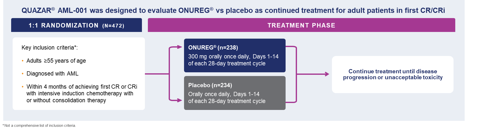
A total of 472 patients were randomized 1:1 to receive either ONUREG® 300 mg or placebo orally on Days 1 through 14 of each 28-day cycle.8 Baseline demographics and disease-related characteristics were well balanced between the ONUREG and placebo arms.8 Across both arms, 72% of patients were 65 years or older, and most patients (92%) had an ECOG PS of 0 or 1. Additionally, approximately three-quarters of patients received 1 or 2 cycles of consolidation therapy.8

With a >2-year median overall survival and a statistically significant survival benefit of ~10 months for patients with AML in first remission compared to placebo, ONUREG met its primary endpoint (24.7 months in the treated arm vs 14.8 months in the placebo arm, hazard ratio (HR) 0.69, 95% confidence interval (CI): 0.55-0.86; P=0.0009).8

The most common adverse reactions (ARs, ≥ 10%) associated with ONUREG® treatment included nausea, vomiting, diarrhea, fatigue/asthenia, constipation, pneumonia, abdominal pain, arthralgia, decreased appetite, febrile neutropenia, dizziness, and pain in extremity.8 Serious ARs occurred in 15% of patients who received ONUREG®, with select Grade 3/4 ARs shown in the table below.8 Eight percent of patients permanently discontinued ONUREG®, 35% of patients required a treatment interruption due to an AR, and 14% of patients required a dose reduction.8

Quazar-Toxicities
ONUREG® is approved for continued treatment of adult patients with acute myeloid leukemia who achieved first complete remission (CR) or complete remission with incomplete blood count recovery (CRi) following intensive induction chemotherapy and are not able to complete intensive curative therapy.8 ONUREG® is an oral hypomethylating agent, offering a convenient, once-daily dosing that patients can take at home.8 However, it is important to emphasize that ONUREG® should not be substituted for intravenous or subcutaneous azacitidine, because the indications and dosing regimen differ between these formulations.8 As the first and only FDA-approved continued AML treatment for patients in first remission, ONUREG® remains an option for appropriate patients.

IMPORTANT SAFETY INFORMATION
CONTRAINDICATIONS
ONUREG® is contraindicated in patients with known severe hypersensitivity to azacitidine or its components.
WARNINGS AND PRECAUTIONS
Risks of Substitution with Other Azacitidine Products
Due to substantial differences in the pharmacokinetic parameters, the recommended dose and schedule for ONUREG® are different from those for the intravenous or subcutaneous azacitidine products. Treatment of patients using intravenous or subcutaneous azacitidine at the recommended dosage of ONUREG® may result in a fatal adverse reaction. Treatment with ONUREG® at the doses recommended for intravenous or subcutaneous azacitidine may not be effective. Do not substitute ONUREG® for intravenous or subcutaneous azacitidine.
Myelosuppression
New or worsening Grade 3 or 4 neutropenia and thrombocytopenia occurred in 49% and 22% of patients who received ONUREG®. Febrile neutropenia occurred in 12%. A dose reduction was required for 7% and 2% of patients due to neutropenia and thrombocytopenia. Less than 1% of patients discontinued ONUREG® due to either neutropenia or thrombocytopenia. Monitor complete blood counts and modify the dosage as recommended. Provide standard supportive care, including hematopoietic growth factors, if myelosuppression occurs.
Increased Early Mortality in Patients with Myelodysplastic Syndromes (MDS)
In AZA-MDS-003, 216 patients with red blood cell transfusion-dependent anemia and thrombocytopenia due to MDS were randomized to ONUREG® or placebo. 107 received a median of 5 cycles of ONUREG® 300 mg daily for 21 days of a 28-day cycle. Enrollment was discontinued early due to a higher incidence of early fatal and/or serious adverse reactions in the ONUREG® arm compared with placebo. The most frequent fatal adverse reaction was sepsis. Safety and effectiveness of ONUREG® for MDS have not been established. Treatment of MDS with ONUREG® is not recommended outside of controlled trials.
Embryo-Fetal Toxicity
ONUREG® can cause fetal harm when administered to a pregnant woman. Azacitidine caused fetal death and anomalies in pregnant rats via a single intraperitoneal dose less than the recommended human daily dose of oral azacitidine on a mg/m2 basis. Advise pregnant women of the potential risk to a fetus. Advise females of reproductive potential to use effective contraception during treatment with ONUREG® and for at least 6 months after the last dose. Advise males with female partners of reproductive potential to use effective contraception during treatment with ONUREG® and for at least 3 months after the last dose.
ADVERSE REACTIONS
Serious adverse reactions occurred in 15% of patients who received ONUREG®. Serious adverse reactions in ≥2% included pneumonia (8%) and febrile neutropenia (7%). One fatal adverse reaction (sepsis) occurred in a patient who received ONUREG®.
Most common (≥10%) adverse reactions with ONUREG® vs placebo were nausea (65%, 24%), vomiting (60%, 10%), diarrhea (50%, 21%), fatigue/asthenia (44%, 25%), constipation (39%, 24%), pneumonia (27%, 17%), abdominal pain (22%, 13%), arthralgia (14%, 10%), decreased appetite (13%, 6%), febrile neutropenia (12%, 8%), dizziness (11%, 9%), pain in extremity (11%, 5%).
LACTATION
There are no data regarding the presence of azacitidine in human milk or the effects on the breastfed child or milk production. Because of the potential for serious adverse reactions in the breastfed child, advise women not to breastfeed during treatment with ONUREG® and for 1 week after the last dose.

Please see full Prescribing Information for ONUREG®.

References
1. National Cancer Institute. SEER Cancer Statistics Factsheets: Acute Myeloid Leukemia. http://seer.cancer.gov/statfacts/html/amyl.html. Accessed April 21, 2021.
2. Medeiros BC, Satram S. Real world treatment patterns and comparative effectiveness among elderly patients with acute myeloid leukemia in the United States. Ann Hematol Oncol. 2020;7(1):1283.
3. Burnett A, Wetzler M, Löwenberg B. Therapeutic advances in acute myeloid leukemia. J Clin Oncol. 2011;29(5):487-494.
4. Brinda B, Khan I, Parkin B, Konig H. The rocky road to personalized medicine in acute myeloid leukaemia. J Cell Mol Med. 2018;22(3):1411-1427.
5. Lai C, Doucette K, Norsworthy K. Recent drug approvals for acute myeloid leukemia. J Hematol Oncol. 2019;12(100):1-20.
6. Medeiros BC. Interpretation of clinical endpoints in trials of acute myeloid leukemia. Leuk Res. 2018;68:32-29.
7. Medeiros BC, Chan SM, Daver NG, Jonas BA, Pollyea DA. Optimizing survival outcomes with post-remission therapy in acute myeloid leukemia. Am J Hematol. 2019;94:803-811.
8. ONUREG® [Prescribing Information]. Summit, NJ: Celgene Corporation; 2021.

© 2021 Celgene Corporation.
ONUREG is a trademark of Celgene Corporation, a Bristol-Myers Squibb company.
QUAZAR is a registered trademark of Celgene Corporation, a Bristol-Myers Squibb company.
1/22 2011-US-2100199

Overall Survival Benefit with the Addition of Capecitabine to Adjuvant Chemotherapy

SUMMARY: Breast cancer is the most common cancer among women in the US and about 1 in 8 women (12%) will develop invasive breast cancer during their lifetime. Approximately 290,560 new cases of breast cancer will be diagnosed in 2022 and about 43,780 individuals will die of the disease, largely due to metastatic recurrence.

Patients with early stage breast cancer often receive adjuvant chemotherapy to improve Overall Survival (OS), and this is even more so true for HER positive and triple negative (ER, PR and HER negative) breast cancer patients, who are at an increased risk to develop recurrent disease. Meta-analyses conducted by the Early Breast Cancer Trialists’ Collaborative Group (EBCTCG) has shown a 20-25% relative risk reduction in breast cancer mortality with first-generation adjuvant chemotherapy regimens such as CMF (Cyclophosphamide/Methotrexate/Fluorouracil) and additional survival benefit with the Anthracyclines and Taxane based regimens. This benefit is dependent on the type of chemotherapy administered and chemotherapy dose intensity.

XELODA® (Capecitabine) is an oral prodrug of fluorouracil which is presently approved for the treatment of advanced breast cancer, but NOT for neoadjuvant or adjuvant treatment of early breast cancer. Meta-analysis of randomized trials has found that addition of Capecitabine to standard adjuvant chemotherapy regimens prolongs Disease Free Survival (DFS), whereas replacing a standard agent with Capecitabine did not improve DFS. Preclinical models have suggested that chemotherapy agents such as taxanes, and Cyclophosphamide increase thymidine phosphorylase concentration in the cancer cell, potentially leading to improved conversion of Capecitabine to fluorouracil within the tumor, suggesting that concomitant administration of Capecitabine with these agents improves efficacy, compared with single-agent Capecitabine. The researchers in this publication addressed the question whether addition of Capecitabine to these regimens could lead to improved survival outcomes.

The Finland Capecitabine Trial (FinXX) is a randomized, open-label, multicenter, Phase III trial that evaluated the addition of Capecitabine to an adjuvant chemotherapy regimen containing a taxane and an anthracycline for the treatment of early breast cancer. In this study, 1,500 patients with axillary node-positive or high-risk node-negative early breast cancer were randomly assigned to 6 cycles of either the Capecitabine arm- TX-CEX (N=753) or to the control group-T-CEF (N=747). TX-CEF consisted of Docetaxel 60 mg/m2 IV day 1 and Capecitabine 900 mg/m2 PO BID on days 1-15 of a 21-day cycle for 3 cycles followed by CEX consisting of Cyclophosphamide 600 mg/m2 IV Day 1, Epirubicin 75 mg/m2 IV on Day 1 and Capecitabine 900 mg/m2 PO BID on days 1-15 of a 21 day cycle for 3 cycles. T-CEF consisted of Docetaxel 80 mg/m2 IV Day 1, every 3 weeks for 3 cycles followed by CEF consisting of Cyclophosphamide 600 mg/m2 IV, Epirubicin 75 mg/m2 IV and Fluorouracil 600 mg/m2 IV, all administered on Day 1, every 3 weeks for 3 cycles. Adjuvant endocrine therapy was initiated within 2 months after completion of chemotherapy if the tumor was ER or PR-positive. Radiotherapy was given after completion of chemotherapy according to each institution’s practice. Adjuvant Trastuzumab was approved while the trial accrual was ongoing and was allowed for women with HER2-positive cancer after May 2005, and adjuvant Trastuzumab was administered to 13% patients assigned to TX-CEF and 11% patients assigned to T-CEF. The median patient age was 52.5 yrs, 76% of patients had ER-positive tumors, 19% had HER2-positive cancer, 13% had Triple Negative Breast Cancer, more than 90% had T1 or T2 tumors, and 89% were node positive. The researchers then performed a protocol-scheduled analysis of Overall Survival on the basis of approximately 15-year follow up of the patients.

At a median follow up of 15.3 years, the Overall Survival was 77.6% in the TX-CEX group and 73.3% in the T-CEF group (HR=0.81; P=0.037). Exploratory subgroup analysis suggested that patients with ER-negative disease and those with Triple Negative Breast Cancer lived longer with the addition of Capecitabine (TX-CEX regimen), than those treated with T-CEF.

It was concluded that the addition of Capecitabine to a chemotherapy regimen significantly improved Overall Survival at median follow up of 15 years in a patient population with early breast cancer, suggesting that Capecitabine may be an important addition to adjuvant chemotherapy in patients with high risk disease.

Adjuvant Capecitabine for Early Breast Cancer: 15-Year Overall Survival Results from a Randomized Trial. Joensuu H, Kellokumpu-Lehtinen P-L , Huovinen R, et al. DOI: 10.1200/JCO.21.02054 Journal of Clinical Oncology. Published online January 12, 2022.

FDA Approves ENJAYMO® for Cold Agglutinin Disease

SUMMARY: The FDA on February 4, 2022 approved ENJAYMO® (Sutimlimab-jome) to decrease the need for Red Blood Cell transfusion due to hemolysis in adults with Cold Agglutinin Disease (CAD). CAD is a chronic and rare autoimmune hemolytic anemia estimated to impact the lives of 5,000 individuals in the U.S. and accounts for 15% of patients with Auto Immune Hemolytic Anemia. It is twice as common in women compared to men and is characterized by hemolysis triggered by exposure to cold temperatures usually 3-4 degrees C (37-39 degrees F). The median age at symptom onset is about 65 years, whereas the median age at diagnosis is about 72 years. The median survival is approximately 11 years.

Cold Agglutinin Disease is a complement-mediated disease and majority of the cases of CAD are mediated by IgM antibodies, unlike warm Auto Immune Hemolytic Anemia, which is predominantly IgG-driven. The circulating IgM antibodies in the body core are not bound to the RBC surface. However, as blood shifts toward the peripheral circulation and cools, IgM antibodies transiently bind to the RBC membrane resulting in agglutination. This in turn activates the complement cascade, binding C3b to the cell surface. As C3b-coated cells return toward the body core, IgM antibody dissociates, and the C3b-coated RBC are destroyed by receptor-specific macrophages present predominantly in the liver and to a lesser degree in the spleen, resulting in extravascular hemolysis and some element of intravascular hemolysis. The severity of hemolysis depends on the thermal amplitude, rather than the serum concentration of IgM antibodies. CAD can be Primary/Idiopathic or Secondary to different underlying disorders such as infections, lymphoproliferative disorders, immunoproliferative disorders, as well as connective tissue diseases.

Patients with CAD usually present with chronic anemia with hemoglobin levels ranging from 4.5 g/dL to 8 g/dL, and over 50% of patients require transfusion support, and therapy is often considered in 75% of the patients. In addition to hemolysis, clinical manifestations include cold-induced circulatory symptoms such as livedo reticularis, Raynaud disease, acrocyanosis and, rarely, cutaneous necrosis. Hemoglobinuria and Splenomegaly tends to be less severe. There has been no approved treatment for CAD. Therapies have included corticosteroids, alkylating agents, purine nucleoside analogs and Rituximab, with little benefit.

ENJAYMO® is a first-in-class humanized monoclonal antibody that is designed to selectively target and inhibit C1 complex in the classical complement pathway, which is part of the innate immune system. By blocking C1 complex, ENJAYMO® inhibits C1-activated hemolysis in CAD and prevents the premature destruction of normal RBC. ENJAYMO® does not inhibit the Lectin and alternative complement pathways.

CARDINAL study is an open-label, single-arm, multicenter trial conducted to assess the efficacy and safety of ENJAYMO® in patients with confirmed CAD and a recent history of RBC transfusion. In this study, eligible patients with CAD (N=24) entered Part A of the study. This consisted of a 26-week treatment period during which patients received ENJAYMO® at a dose of 6.5 g IV for patients weighing less than 75 kg, and 7.5 g IV for those weighing 75 kg or more, on days 0 and 7, after which they received an infusion every 2 weeks. After the 26-week treatment period, patients were eligible to continue to the open-label extension study (Part B). The mean patient age was 71.3 yrs and two thirds of patients had failed prior therapies. CAD diagnosis was defined as the presence of chronic hemolysis, a positive polyspecific Direct Antiglobulin Test result, a monospecific Direct Antiglobulin Test result strongly positive for C3d, a cold agglutinin titer of 1:64 or higher measured at 4°C, a Direct Antiglobulin Test result for IgG of 1+ or less, and no overt malignant disease. Patients with a diagnosis of Systemic Lupus Erythematosus (SLE) or other autoimmune disorder with antinuclear antibodies at screening, overt malignant disease, treatment with Rituximab monotherapy within 3 months before enrollment or treatment with Rituximab combined with chemotherapy within 6 months before enrollment were excluded.

The Primary end point was a composite of a normalization of the hemoglobin level to 12 g or more per deciliter or an increase in the hemoglobin level of 2 g or more per deciliter from baseline, without RBC transfusion from week 5 through week 26. Secondary end points included improvements in hemoglobin level, normalization of bilirubin and LDH level, and Quality of Life, as assessed with the use of the Functional Assessment of Chronic Illness Therapy (FACIT) Fatigue Scale (scores range from 0 to 52, with a higher score indicating less fatigue. The authors herein reported the results of Part A of the study.

The majority of patients (54%) met the prespecified Primary endpoint criteria with 63% of patients achieving a hemoglobin12 g/dL or more or an increase of at least 2 g/dL and 71% of patients remained transfusion-free after week five. Approximately 92% of patients did not use other therapies for CAD.

With regard to Secondary endpoints, there was a mean increase in the hemoglobin level of 2.29 g/dL at week 3, and 3.18 g/dL at the 26-week treatment assessment time point, from the mean baseline level of 8.6 g/dL. The mean bilirubin levels normalized by week 3, from a mean baseline level of 3.23 mg/dL and clinically meaningful reductions in fatigue were observed by week 1 and were maintained throughout the study. The activity in the classic complement pathway was rapidly inhibited, as assessed by a functional assay. Increased hemoglobin levels, reduced bilirubin levels, and reduced fatigue coincided with inhibition of the classic complement pathway. There were no cases of meningococcal infections and none of the patients experienced thromboembolism.

It was concluded that in this pivotal study, ENJAYMO® had a rapid and sustained treatment benefit in most patients with Cold Agglutinin Disease by preventing chronic hemolysis, markedly increasing hemoglobin levels, and improving quality of life.

Sutimlimab in Cold Agglutinin Disease. Röth A, Barcellini W, D’Sa S, et al. N Engl J Med 2021;384:1323-1334

Consolidation and Maintenance in Newly Diagnosed Multiple Myeloma

SUMMARY: Multiple Myeloma is a clonal disorder of plasma cells in the bone marrow and the American Cancer Society estimates that in the United States, 34,470 new cases will be diagnosed in 2022 and 12,640 patients will die of the disease. Multiple Myeloma is a disease of the elderly, with a median age at diagnosis of 69 years and characterized by intrinsic clonal heterogeneity. Almost all patients eventually will relapse, and patients with a high-risk cytogenetic profile, extramedullary disease or refractory disease have the worst outcomes. The introduction of Proteasome Inhibitors, immunomodulatory agents and CD 38 targeted therapies has resulted in higher Response Rates, as well as longer Progression Free Survival (PFS) and Overall Survival (OS), with the median survival for patients with myeloma approaching 10 years or more. Nonetheless, Multiple Myeloma (MM) in 2022 remains an incurable disease.

High Dose Melphalan followed by Autologous Stem Cell Transplantation (HDM/ASCT) remains an important treatment option for transplant eligible patients. REVLIMID® (Lenalidomide) was approved by the FDA in 2017 as maintenance therapy for patients with multiple myeloma following Autologous Stem Cell Transplant (ASCT) and to date is the only drug approved for this indication. Maintenance or Continuous Treatment in patients with newly diagnosed multiple myeloma following induction treatment can result in significantly longer PFS and OS, compared to those patients who receive therapy for a fixed duration of time.

The role of consolidation treatment for newly diagnosed, transplant-eligible patients with multiple myeloma has not been conclusively established and needed prospective evaluation. The present prospective clinical trial was conducted to study the relevance of consolidation therapy using Bortezomib, Lenalidomide, and Dexamethasone (VRD) followed by Lenalidomide maintenance, compared with Lenalidomide maintenance alone, in transplant-eligible newly diagnosed multiple myeloma patients.

The EMN02/HOVON95 trial is an open-label, Phase III study, performed by the European Myeloma Network (EMN) in which 1197 previously untreated transplant-eligible patients with symptomatic Stages I-III Multiple Myeloma were randomly assigned initially to four cycles of Bortezomib, Melphalan, and Prednisone (VMP) or High-Dose Melphalan followed by Autologous Stem Cell Transplantation (HDM/ASCT). Within 2 months after ASCT or last VMP treatment, 878 eligible patients underwent a second randomization to either two 28-day cycles of VRD consolidation treatment, which consisted of Bortezomib 1.3 mg/m2 either IV or SC once daily on days 1, 4, 8, and 11, combined with Lenalidomide 25 mg orally once daily, days 1-21 and Dexamethasone 20 mg orally once daily, on days 1, 2, 4, 5, 8, 9, 11, and 12 (N=451 – Arm B) or no consolidation (N=427 – Arm A). Patients then received Lenalidomide maintenance 10 mg orally once daily on days 1-21 of a 28-day cycle, starting 1-2 months after ASCT or consolidation, and treatment was continued until disease progression or toxicity. The Primary end point was Progression Free Survival (PFS) defined as time from second randomization to disease progression or death. Secondary end points included Partial Response or higher, Overall Survival (OS) from second randomization until death from any cause, and toxicity. Predefined high-risk prognostic subgroups for PFS were cytogenetic abnormalities defined by FISH and included deletion (17p) in 20% or more of enriched plasma cells, t(4;14) in 10% or more of enriched plasma cells, t(14;16) in 10% or more of enriched plasma cells; and amplification 1q. This was in addition to the standard clinical variables such as Hemoglobin level, Serum Creatinine and LDH. Disease assessment was performed before and after consolidation and every 2 months until progression. Bone marrow Minimal Residual Disease (MRD) assessment was performed by multicolor flow cytometry in bone marrow with a detection of 10−4 to 10−5.

At a median follow-up of 74.8 months after the second randomization, the median PFS was significantly prolonged in the VRD consolidation group, compared those who did not receive consolidation treatment (59.3 versus 42.9 months, HR=0.81; P=0.016). This PFS benefit was observed across most predefined subgroups, including Stage, standard-risk cytogenetics, and prior treatment groups. VRD consolidation was however not beneficial in patients with del(17p). Stage III disease and addition of chromosome 1q by FISH at diagnosis were significant adverse prognostic factors for PFS from second randomization. The median duration of maintenance treatment with Lenalidomide was 33 months. Complete Response (CR) or more after consolidation versus no consolidation, and before start of maintenance treatment was 34% versus 18%, respectively (P<0.001). Complete Response or more on protocol including maintenance treatment was 59% with consolidation and 46% without consolidation (P<0.001). Minimal Residual Disease analysis in a subgroup of 226 VRD-consolidated patients with CR or stringent CR or Very Good Partial Response before start of maintenance treatment demonstrated a 74% MRD-negativity rate. Toxicities related to VRD consolidation were acceptable and manageable.

It was concluded from this study that consolidation followed by maintenance treatment after either Bortezomib, Melphalan and Prednisone or High-Dose Melphalan and Autologous Stem Cell Transplantation, significantly improves Progression Free Survival and Overall Response Rate in transplant-eligible and Lenalidomide-naïve newly diagnosed patients with Multiple Myeloma, compared to maintenance treatment alone.

Consolidation and Maintenance in Newly Diagnosed Multiple Myeloma. Sonneveld P, Dimopoulos MA, Beksac M, et al. J Clin Oncol. 2021;39:3613-3622.

LAG-3 Inhibitor Relatlimab and OPVIDO® in Advanced Untreated Melanoma

SUMMARY: The American Cancer Society’s estimates that for 2022, about 99,780 new cases of melanoma of the skin will be diagnosed in the United States and 7,650 people are expected to die of the disease. The rates of melanoma have been rising rapidly over the past few decades, but this has varied by age.

A better understanding of Immune checkpoints has opened the doors for the discovery of novel immune targets. Immune checkpoints are cell surface inhibitory proteins/receptors that harness the immune system and prevent uncontrolled immune reactions. Survival of cancer cells in the human body may be related to their ability to escape immune surveillance, by inhibiting T lymphocyte activation. Under normal circumstances, inhibition of an intense immune response and switching off the T cells of the immune system is accomplished by Immune checkpoints or gate keepers. With the recognition of Immune checkpoint proteins and their role in suppressing antitumor immunity, antibodies have been developed that target the membrane bound inhibitory Immune checkpoint proteins/receptors such as CTLA-4 (Cytotoxic T-Lymphocyte Antigen 4, also known as CD152), PD-1(Programmed cell Death 1), etc. By blocking the Immune checkpoint proteins, T cells are unleashed, resulting in T cell proliferation, activation and a therapeutic response.

YERVOY® (Ipilimumab), a fully human immunoglobulin G1 monoclonal antibody that blocks Immune checkpoint protein/receptor CTLA-4 was compared with PD-1 inhibitors, OPDIVO® (Nivolumab) and KEYTRUDA® (Pembrolizumab) in patients with advanced melanoma, and both OPDIVO® and KEYTRUDA® have demonstrated superior Overall Survival (OS), Progression Free Survival (PFS), and Objective Response Rate (ORR), and with a better safety profile. In the CheckMate 067, which is a double-blind Phase III study, results from the 6.5 year analysis showed that a combination of OPDIVO® plus YERVOY® demonstrated significant improvement in OS and PFS, when compared to single agent OPDIVO® or single agent YERVOY®.

In an attempt to improve outcomes and enhance the risk-benefit profiles of immunotherapy combinations, alternate Immune checkpoints are being explored. LAG-3 (Lymphocyte-Activation Gene 3 (LAG-3), is a cell-surface receptor expressed on immune cells including activated CD4+ T cells, and negatively regulates T-cell proliferation, inhibits T-cell activation and effector T-cell function. LAG-3 is upregulated in several tumor types, including malignant melanoma.

Relatlimab is a first-in-class human IgG4 LAG-3–blocking antibody that binds to LAG-3 and restores the effector function of exhausted T cells, resulting in T cell proliferation, activation and a therapeutic response. In preclinical studies, dual inhibition of LAG-3 and PD-1 showed synergistic antitumor activity, and in a Phase I/II trial, the combination of Relatlimab and Nivolumab, demonstrated durable Objective Responses in patients with Relapsed/Refractory melanoma following treatment with PD-1 inhibitors.

RELATIVITY-047 is a Phase II/III, global, multicenter, double-blind, randomized trial in which a fixed-dose combination of Relatlimab and Nivolumab was compared with Nivolumab alone, in patients with previously untreated metastatic or unresectable melanoma. In this study, 714 patients were randomly assigned 1:1 to receive Relatlimab 160 mg and Nivolumab 480 mg in a fixed-dose combination (N=355) or single agent Nivolumab 480 mg (N=359). Both regimens were administered as an IV infusion over 60 minutes every 4 weeks, and treatment was continued until disease progression, unacceptable toxicities, or withdrawal of consent. Both treatment groups were well balanced and patients were stratified according to LAG-3 expression (1% or more versus less than 1%), PD-L1 expression (1% or more versus less than 1%), BRAF V600 mutation status, and metastasis stage (M0 or M1 with normal LDH levels versus M1 with elevated LDH levels). More patients in the Relatlimab-Nivolumab group had Stage M1c disease, and a larger proportion had three or more sites with at least one metastatic lesion. The Primary end point was Progression Free Survival (PFS) as assessed by blinded Independent Central Review. Secondary end points included Overall Survival and Objective Response Rate (ORR). The median follow up was 13.2 months and the use of subsequent therapies upon progression was similar in the two treatment groups.

The median PFS was 10.1 months with Relatlimab-Nivolumab as compared with 4.6 months with Nivolumab (HR=0.75; P=0.006). The PFS benefit at 12 months with Relatlimab-Nivolumab was 47.7% compared to 36.0% with Nivolumab. The PFS benefit was more so with Relatlimab- Nivolumab across key prespecified subgroups, compared to single agent Nivolumab. Patients with poor prognosis characteristics, such as visceral metastases, high tumor burden, elevated levels of serum LDH, or mucosal or acral melanoma, had better outcomes with Relatlimab-Nivolumab combination, than with single agent Nivolumab. Further, a benefit with Relatlimab-Nivolumab was also noted across BRAF mutant and wild-type subgroups, compared to Nivolumab. Expression of LAG-3 or PD-L1 was not useful in predicting a benefit of Relatlimab-Nivolumab over single agent Nivolumab and appears to NOT have a clear role in treatment selection.

Grade 3 or 4 toxicities occurred in 18.9% of patients in the Relatlimab-Nivolumab group and in 9.7% of patients in the single agent Nivolumab group. The Safety profile of Relatlimab-Nivolumab appeared favorable, when compared with dual checkpoint inhibition with a CTLA-4 inhibitor and PD-1 inhibitor combination (Ipilimumab-Nivolumab) in the CheckMate 067 trial, in which Adverse Events were noted in 59% of patients.

It was concluded that inhibition of two immune checkpoints, LAG-3 and PD-1, provided greater benefit with regards to Progression Free Survival, than inhibition of PD-1 alone, in patients with previously untreated metastatic or unresectable melanoma. The authors added that these results validate blocking LAG-3 in combination with PD-1 as a therapeutic strategy for patients with melanoma, and establishes LAG-3 as the third immune checkpoint pathway, thus providing more treatment options for patients with advanced melanoma.

Relatlimab and Nivolumab versus Nivolumab in Untreated Advanced Melanoma. Tawbi HA, Schadendorf D, Lipson EJ, et al. for the RELATIVITY-047 Investigators. N Engl J Med 2022;386:24-34.