FDA Approves KEYTRUDA® Plus Chemotherapy for Triple Negative Breast Cancer

SUMMARY: The FDA on November 13, 2020, granted accelerated approval to KEYTRUDA® (Pembrolizumab) in combination with chemotherapy for the treatment of patients with locally recurrent, unresectable or metastatic, Triple Negative Breast Cancer (TNBC), whose tumors express PD-L1 (Combined Positive Score-CPS 10 or more) as determined by an FDA approved test. Breast cancer is the most common cancer among women in the US and about 1 in 8 women (13%) will develop invasive breast cancer during their lifetime. Approximately 276,480 new cases of invasive female breast cancer will be diagnosed in 2020 and about 42,170 women will die of the disease. Triple Negative Breast Cancer (TNBC) is a heterogeneous, molecularly diverse group of breast cancers and are ER (Estrogen Receptor), PR (Progesterone Receptor) and HER2 (Human Epidermal Growth Factor Receptor-2) negative. TNBC accounts for 15-20% of invasive breast cancers, with a higher incidence noted in young patients and African American females. It is usually aggressive, and tumors tend to be high grade, and patients with TNBC are at a higher risk of both local and distant recurrence and often develop visceral metastases. Those with metastatic disease have one of the worst prognoses of all cancers with a median Overall Survival of 13 months. The majority of patients with TNBC who develop metastatic disease do so within the first 3 years after diagnosis, whereas those without recurrence during this period of time have survival rates, similar to those with ER-positive breast cancers. The lack of known recurrent oncogenic drivers in patients with metastatic TNBC, presents a major therapeutic challenge. Overall survival among patients with pretreated metastatic TNBC has not changed over the past 2 decades and standard chemotherapy is associated with low response rates of 10-15% and a Progression Free Survival (PFS) of only 2-3 months.Unleashing-T-Cell-Function-with-Immune-Checkpoint-Inhibitors

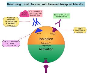
KEYTRUDA® is a fully humanized, Immunoglobulin G4, anti-PD-1, monoclonal antibody, that binds to the PD-1 receptor and blocks its interaction with ligands PD-L1 and PD-L2. It thereby reverses the PD-1 pathway-mediated inhibition of the immune response, and unleashes the tumor-specific effector T cells. The rationale for combining chemotherapy with immunotherapy is that cytotoxic chemotherapy releases tumor-specific antigens, and immune checkpoint inhibitors such as KEYTRUDA® when given along with chemotherapy can enhance endogenous anticancer immunity. Single agent KEYTRUDA® in metastatic TNBC demonstrated durable antitumor activity in several studies, with Objective Response Rates (ORRs) ranging from 10% to 21% and improved clinical responses in patients with higher PD-L1 expression. When given along with chemotherapy as a neoadjuvant treatment for patients with high-risk, early-stage TNBC, KEYTRUDA® combination achieved Pathological Complete Response rate of 65%, regardless of PD-L1 expression. Based on this data, KEYTRUDA® in combination with chemotherapy was studied, for first-line treatment of TNBC.

KEYNOTE-355 is a randomized, double-blind, Phase III study, which evaluated the benefit of KEYTRUDA® in combination with one of the three different chemotherapy regimens, nab-Paclitaxel, Paclitaxel, or the non-taxane containing Gemzar/Carboplatin, versus placebo plus one of the three chemotherapy regimens, in patients with previously untreated or locally recurrent inoperable metastatic TNBC. In this study, 847 patients were randomized 2:1 to receive either KEYTRUDA® 200 mg IV on day 1 of each 21-day cycle along with either nab-paclitaxel 100 mg/m2 IV on days 1, 8 and 15 of each 28-day cycle, Paclitaxel 90 mg/m2 IV on days 1, 8 and 15 of each 28-day cycle, or Gemcitabine 1000 mg/m2 IV plus Carboplatin AUC 2, IV on days 1 and 8 of each 21-day cycle (N= 566) or placebo along with one of the three chemotherapy regimens (N= 281). This study was not designed to compare the efficacy of the different chemotherapy regimens. Treatment was continued until disease progression. Patients were stratified by chemotherapy, PD-L1 tumor expression (CPS of 1 or higher versus CPS of less than 1), and prior treatment with the same class of neoadjuvant/adjuvant chemotherapy (Yes versus No). The baseline characteristics of treatment groups were well-balanced. The co-Primary end points of the trial were Progression Free Survival (PFS) and Overall Survival (OS) in patients with PD-L1-positive tumors, and in all patients. Secondary end points were Objective Response Rate (ORR), Duration of Response, Disease Control Rate, and Safety. The median follow up for patients assigned to receive KEYTRUDA® was 17.5 months and 15.5 months for the placebo group. The authors reported the results from an interim analysis conducted by an Independent Data Monitoring Committee (IDMC).

KEYTRUDA® in combination with chemotherapy, significantly improved PFS in patients with CPS (Combined Positive Score) of 10 or greater. The median PFS was 9.7 months for KEYTRUDA® plus chemotherapy, compared with 5.6 months for placebo plus chemotherapy (HR=0.65, P=0.0012). This represented a 35% reduction in the risk of disease progression. Among patients with CPS of 1 or greater, the median PFS was 7.6 months for KEYTRUDA® plus chemotherapy, compared with 5.6 months for the placebo plus chemotherapy arm (HR= 0.74; P=0.0014). This however based on prespecified statistical criteria, was not considered statistically significant. Among the entire Intention-To-Treat (ITT) population, the median PFS was 7.5 months in the KEYTRUDA® plus chemotherapy group, compared with 5.6 months for chemotherapy plus placebo group (HR=0.82). Formal statistical significance was not tested in the ITT population. Overall Survival data are pending. Adverse Events (AEs) were similar in both treatment groups, although immune-related AEs occurred at a higher incidence in the KEYTRUDA® arm.

It was concluded that KEYTRUDA® in combination with several chemotherapy regimens, showed a statistically significant and clinically meaningful improvement in PFS, compared with chemotherapy alone, in patients with previously untreated locally recurrent, inoperable or metastatic TNBC, whose tumors expressed PD-L1 with a Combined Positive Score (CPS) of 10 or more. This data may be particularly relevant for patients who may have received a taxane in the adjuvant setting within a year, and could be more appropriately treated with a non-taxane regimen, in combination with KEYTRUDA®.

KEYNOTE-355: Randomized, double-blind, phase III study of pembrolizumab + chemotherapy versus placebo + chemotherapy for previously untreated locally recurrent inoperable or metastatic triple-negative breast cancer. Cortes J, Cescon DW, Rugo HS. et al. J Clin Oncol 38: 2020 (suppl; abstr 1000)

RUBRACA® in Metastatic Castrate Resistant Prostate Cancer with BRCA Mutations

SUMMARY: Prostate cancer is the most common cancer in American men with the exclusion of skin cancer, and 1 in 9 men will be diagnosed with Prostate cancer during their lifetime. It is estimated that in the United States, about 191,930 new cases of Prostate cancer will be diagnosed in 2020 and 33,330 men will die of the disease.

The development and progression of Prostate cancer is driven by androgens. Androgen Deprivation Therapy (ADT) or testosterone suppression has therefore been the cornerstone of treatment of advanced Prostate cancer and is the first treatment intervention. Androgen Deprivation Therapies have included bilateral orchiectomy or Gonadotropin Releasing Hormone (GnRH) analogues, with or without first generation Androgen Receptor (AR) inhibitors such as CASODEX® (Bicalutamide), NILANDRON® (Nilutamide) and EULEXIN® (Flutamide) or with second-generation, anti-androgen agents, which include, ZYTIGA® (Abiraterone), XTANDI® (Enzalutamide) and ERLEADA® (Apalutamide). Approximately 10-20% of patients with advanced Prostate cancer will progress to Castration Resistant Prostate Cancer (CRPC) within five years during ADT, and over 80% of these patients will have metastatic disease at the time of CRPC diagnosis. The estimated mean survival of patients with CRPC is 9-36 months, and there is therefore an unmet need for new effective therapies.

DNA damage is a common occurrence in daily life by UV light, ionizing radiation, replication errors, chemical agents, etc. This can result in single and double strand breaks in the DNA structure which must be repaired for cell survival. The two vital pathways for DNA repair in a normal cell are BRCA1/BRCA2 and PARP. BRCA1 and BRCA2 genes recognize and repair double strand DNA breaks via Homologous Recombination Repair (HRR) pathway. Homologous Recombination is a type of genetic recombination, and is a DNA repair pathway utilized by cells to accurately repair DNA double-stranded breaks during the S and G2 phases of the cell cycle, and thereby maintain genomic integrity. Homologous Recombination Deficiency (HRD) is noted following mutation of genes involved in HR repair pathway. At least 15 genes are involved in the Homologous Recombination Repair (HRR) pathway including BRCA1, BRCA2 and ATM genes. The BRCA1 gene is located on the long (q) arm of chromosome 17 whereas BRCA2 is located on the long arm of chromosome 13. BRCA1 and BRCA2 are tumor suppressor genes and functional BRCA proteins repair damaged DNA, and play an important role in maintaining cellular genetic integrity. They regulate cell growth and prevent abnormal cell division and development of malignancy.

Recently published data has shown that deleterious Germline and/or Somatic mutations in BRCA1, BRCA2, ATM, or other Homologous Recombination DNA-repair genes, are present in about 25% of patients with advanced prostate cancer, including mCRPC. Approximately 12% of men with mCRPC harbor a deleterious BRCA1 or BRCA2 mutation (BRCA1, 2%; BRCA2, 10%). Mutations in BRCA1 and BRCA2 also account for about 20-25% of hereditary breast cancers, about 5-10% of all breast cancers, and 15% of ovarian cancers. BRCA mutations can either be inherited (Germline) and present in all individual cells or can be acquired and occur exclusively in the tumor cells (Somatic). Somatic mutations account for a significant portion of overall BRCA1 and BRCA2 aberrations. Loss of BRCA function due to frequent somatic aberrations likely deregulates HR pathway, and other pathways then come in to play, which are less precise and error prone, resulting in the accumulation of additional mutations and chromosomal instability in the cell, with subsequent malignant transformation. HRD therefore indicates an important loss of DNA repair function. The PARP (Poly ADP Ribose Polymerase), family of enzymes include, PARP1and PARP2, and is a related enzymatic pathway that repairs single strand breaks in DNA. In a BRCA mutant, the cancer cell relies solely on PARP pathway for DNA repair to survive. PARP inhibitors trap PARP onto DNA at sites of single-strand breaks, preventing their repair and generating double-strand breaks that cannot be repaired accurately in tumors harboring defects in Homologous Recombination Repair pathway genes, such as BRCA1 or BRCA2 mutations, and this leads to cumulative DNA damage and tumor cell death.MOA-of-RUBRACA

RUBRACA® (Rucaparib) is an oral, small molecule inhibitor of PARP. TRITON2 is an international, multicenter, open-label, single arm, Phase II trial, in which patients with BRCA-mutated mCRPC, who had progressed after one to two lines of next-generation Androgen Receptor-directed therapy and one taxane-based chemotherapy for mCRPC were included. In this study, 115 mCRPC patients with either Germline or Somatic BRCA mutations, with or without measurable disease were enrolled, of whom 62 patients (54%) had measurable disease at baseline. Patients received RUBRACA® 600 mg orally twice daily and concomitant GnRH analog or had prior bilateral orchiectomy. Treatment was continued until disease progression or unacceptable toxicity. The median patient age was 72 years, majority of patients had an ECOG performance status of 0 or 1, 67% of patients had Gleason score of 8 or more at diagnosis, 68% had bone-only disease and 47% had 10 or more bone lesions. The Primary endpoint was Objective Response Rate (ORR) by blinded IRR (Independent Radiology Review), as well as ORR by investigator assessment. Secondary end points included Duration of Response (DOR) in those with measurable disease, locally assessed PSA response rate (50% or more decrease from baseline) rate, Overall Survival (OS), and Safety. The median follow up was 17.1 months.

The confirmed ORR for the IRR-evaluable population was 43.5%, and the confirmed ORR for the investigator-evaluable population was 50.8%. The median DOR was not evaluable and 56% of patients with confirmed Objective Responses had a DOR of 6 months or more. The confirmed PSA response rate was 54.8% and the median time to PSA response was 1.9 months. The Objective Response Rates were similar for patients with a Germline or Somatic BRCA mutations, and for patients with a BRCA1 or BRCA2 mutations. However, a higher PSA response rate was observed in patients with a BRCA2 mutation. The median radiographic Progression Free Survival was 9.0 months per IRR assessment and 8.5 months per investigator assessment. The OS data were not yet mature at the time of the analysis. The most frequent Grade 3 or more treatment related Adverse Event was anemia (25.2%).

It was concluded that RUBRACA® demonstrates promising efficacy in patients with mCRPC with deleterious BRCA mutations. TRITON3 study is evaluating RUBRACA® versus physician’s choice of second-line AR-directed therapy or Docetaxel, in chemotherapy-naïve patients with mCRPC and alterations in BRCA1/2, who progressed on one prior AR-directed therapy.

Rucaparib in Men With Metastatic Castration-Resistant Prostate Cancer Harboring a BRCA1 or BRCA2 Gene Alteration. Abida W, Patnaik A, Campbell D, et al. on behalf of the TRITON2 investigators. J Clin Oncol. 2020;38:3763-3772.

NERLYNX® Combination Superior to TYKERB® Combination in Advanced HER2-Positive Breast Cancer

SUMMARY: Breast cancer is the most common cancer among women in the US and about 1 in 8 women (13%) will develop invasive breast cancer during their lifetime. Approximately 276,480 new cases of invasive female breast cancer will be diagnosed in 2020 and about 42,170 women will die of the disease. The HER or erbB family of receptors consist of HER1, HER2, HER3 and HER4. Approximately 15-20% of invasive breast cancers overexpress HER2/neu oncogene, which is a negative predictor of outcomes without systemic therapy. Patients with HER2-positive metastatic breast cancer are often treated with anti-HER2 targeted therapy along with chemotherapy, irrespective of hormone receptor status, and this has resulted in significantly improved treatment outcomes.HER2-Directed-Therapy

NERLYNX® (Neratinib) is a potent, irreversible, oral Tyrosine Kinase Inhibitor, of HER1, HER2 and HER4 (pan-HER inhibitor). NERLYNX® interacts with the catalytic domain of HER1, HER2, and HER4 and blocks their downstream signaling pathways, resulting in decreased cell proliferation and increased cell death. Clinical data has suggested that NERLYNX® has significant activity in suppressing HER-mediated tumor growth and is able to overcome tumor escape mechanisms experienced with current HER2-targeted and chemotherapeutic agents. It has been well known that hormone receptor positive breast cancer patients, who are also HER2-positive, have relative resistance to hormone therapy. Preclinical models had suggested that the addition of NERLYNX® could improve responses in ER positive, HER2-positive breast cancer patients. Further, NERLYNX® has clinical activity in patients with HER2-positive metastatic breast cancer. NERLYNX® is the first TKI approved by the FDA, shown to reduce the risk for disease recurrence, in patients with early stage HER2-positive breast cancer. NERLYNX® when given for 12 months after chemotherapy and HERCEPTIN®-based adjuvant therapy, to women with HER2-positive breast cancer, significantly reduced the proportion of clinically relevant breast cancer relapses that might lead to death, such as distant and locoregional recurrences outside the preserved breast.

TYKERB® (Lapatinib) is a Tyrosine Kinase Inhibitor of HER2 and EGFR, and in a previously published Phase III study, a combination of TYKERB® plus XELODA® (Capecitabine) was found to be superior to XELODA® alone in women with HER2-positive advanced breast cancer, that has progressed after treatment with regimens that included an Anthracycline, a Taxane, and HERCEPTIN®. (N Engl J Med 2006; 355:2733-2743)

The NALA trial was designed to compare NERLYNX® plus XELODA® versus TYKERB® plus XELODA® in patients with heavily pretreated Stage IV HER2-positive metastatic breast cancer, including those with asymptomatic or stable (treated or untreated) CNS metastases. In this multinational, randomized, active-controlled, Phase III study, 621 patients (N = 621) with metastatic HER2-positive breast cancer who received two or more prior anti-HER2 based regimens in the metastatic setting were randomly assigned in a 1:1 to receive NERLYNX® 240 mg given orally once daily on days 1-21 along with XELODA® 750 mg/m2 given orally twice daily on days 1-14 for each 21-day cycle (N=307) or TYKERB® 1250 mg given orally once daily on days 1-21 along with XELODA® 1000 mg/m2 given orally twice daily on days 1-14 for each 21-day cycle (N=314). Approximately 85% of patients had visceral metastases, and about 30% had received at least three anti-HER2 therapies. Patients in the NERLYNX® group also received antidiarrheal prophylaxis with Loperamide. Patients were treated until disease progression or unacceptable toxicity. The Co-Primary endpoints were Progression Free Survival (PFS) and Overall Survival (OS). Secondary endpoints included Objective Response Rate (ORR) and Duration of Response, Clinical Benefit Rate (CBR), time to intervention for symptomatic metastatic Central Nervous System (CNS) disease and Safety.

At a median follow up of 29.9 months, treatment with NERLYNX® with XELODA® significantly improved the median PFS, compared to those receiving TYKERB® with XELODA® (HR=0.76; P=0.006). This represented a 24% reduction in the risk of disease progression or death for those receiving a combination of NERLYNX® and XELODA®. The PFS rate at 12 months was 29% versus 15% respectively. The median OS was 21 months for patients receiving NERLYNX® and XELODA® compared to 18.7 months for those receiving TYKERB® and XELODA® (HR=0.88; P=0.20) and this was not statistically significant. The ORR was numerically higher with NERLYNX® and XELODA® combination in patients with measurable disease (32.8% versus 26.7%), and there was a statistically significant improvement in the Clinical Benefit Rate (45% versus 36%; P=0.03). The median Duration of Response was 8.5 versus 5.6 months respectively (HR=0.50; P=0.0004), favoring the NERLYNX® combination. The time to intervention for symptomatic CNS disease was significantly delayed with NERLYNX® combination versus TYKERB® combination, with an overall cumulative incidence of 22.8% versus 29.2% respectively (P= 0.043). The most common toxicities of any grade in the study population were diarrhea, nausea, palmar-plantar erythrodysesthesia syndrome, and vomiting. Treatment related toxicities were similar between arms, but there was a higher rate of Grade 3 diarrhea with the NERLYNX® combination (24% versus 13% respectively).

It was concluded from this study that a combination of NERLYNX® and XELODA® significantly improved Progression Free Survival, with a trend towards improved Overall Survival, and also resulted in a delayed time to intervention for symptomatic CNS disease, among patients with heavily pretreated advanced HER2-positive breast cancer. This is the first study to demonstrate superiority of one HER2-directed Tyrosine Kinase Inhibitor over another, in HER2-positive metastatic breast cancer.

Neratinib Plus Capecitabine Versus Lapatinib Plus Capecitabine in HER2-Positive Metastatic Breast Cancer Previously Treated With 2 or More HER2-Directed Regimens: Phase III NALA Trial. Saura C, Oliveira M, Y Feng Y-H, et al. for the NALA Investigators. J Clin Oncol. 2020;38:3138-3149.

Chemotherapy-Free First Line Induction and Consolidation Treatment for Acute Lymphocytic Leukemia

SUMMARY: It is estimated that 6150 individuals will be diagnosed with Acute Lymphocytic Leukemia (ALL) in the US and 1520 patients will die of the disease. ALL is more common in children, but can occur at any age and arises from malignant transformation of B- or T-cell progenitor cells. These cells express surface antigens that define their respective lineages. Precursor B-cell ALL cells typically express CD10, CD19, and CD34 on their surface, along with nuclear Terminal deoxynucleotide Transferase (TdT), whereas precursor T-cell ALL cells commonly express CD2, CD3, CD7, CD34, and TdT.

Philadelphia Chromosome (Chromosome 22) is a result of a reciprocal translocation between chromosomes 9 and 22, wherein the ABL gene from chromosome 9 fuses with the BCR gene on chromosome 22. As a result, the auto inhibitory function of the ABL gene is lost and the BCR-ABL fusion gene is activated resulting in cell proliferation and leukemic transformation of hematopoietic stem cells. Approximately 20% of adults and a small percentage of children with ALL are Philadelphia Chromosome (Ph) positive, and in the majority of children and in more than 50% of adults with Ph-positive ALL, the molecular abnormality (fusion protein) is different from that in Ph-positive Chronic Myelogenous Leukemia (p190 versus p210).

Adult patients with Ph-positive ALL are rarely cured with chemotherapy and the prognosis in these patients has markedly improved with the availability of BCR/ABL targeted Tyrosine Kinase Inhibitors (TKIs). Use of these TKIs with or without chemotherapy can result in a Complete Hematologic Remission in 94-100% of patients, irrespective of age. Eligible patients are then usually referred for allogeneic Hematopoietic Stem Cell Transplant (allo HSCT). To increase the chance of cure and decrease the likelihood of relapse, sustained decrease in Minimal Residual Disease is required, with a reduction in the tumor burden to less than 1 tumor cell in 10,000 bone marrow mononuclear cells.BLINATUMOMAB-(BLINCYTO)-(Engages-Two-Different-Targets-Simultaneously)

BiTE® technology (Bispecific T cell Engager antibody) engages the body’s immune system to detect and target malignant cells. These modified antibodies are designed to engage two different targets simultaneously, thereby placing the patient’s T cells within reach of the targeted cancer cell and facilitating apoptosis of the cancer cell. BiTE® antibodies are currently being investigated to treat a wide variety of malignancies. BLINCYTO® (Blinatumomab) is a BiTE® antibody designed to activate the patients T cells with its anti-CD3 group and then bind them to tumor cells with its anti-CD19 group, thus promoting cellular cytotoxicity. CD19 is a protein expressed on the surface of B-cell derived leukemias and lymphomas

The Italian GIMEMA investigators adopted a chemotherapy-free induction strategy and conducted a Phase II single-group trial, in which adults (no upper age limit) with newly diagnosed Ph-positive ALL, received first line therapy with SPRYCEL® (Dasatinib) plus glucocorticoids, followed by two cycles of BLINCYTO® (Blinatumomab). This study enrolled 63 patients with newly diagnosed Ph-positive ALL, and patients received prephase treatment with a glucocorticoid for 7 days before they received SPRYCEL®, and glucocorticoids were continued for an additional 24 days and discontinued on day 31. SPRYCEL® 140 mg orally once daily was administered as induction therapy for 85 days.

Patients who completed the induction phase received consolidation treatment with BLINCYTO® 28 mcg per day, and before each BLINCYTO® cycle, Dexamethasone 20 mg was administered. A minimum of two cycles of BLINCYTO® was mandatory and up to three additional cycles were allowed. Levetiracetam 500 mg twice daily was administered during treatment with BLINCYTO®, to prevent CNS adverse events. SPRYCEL® was continued during treatment with BLINCYTO®, and after BLINCYTO® administration, except in those patients in whom a T315I mutation was detected during the induction phase. Lumbar punctures were performed at diagnosis, at days 14, 22, 43, 57, and 85, and at the end of each BLINCYTO® cycle, for a total of 12 procedures. The choice of postconsolidation treatment, including allogeneic HSCT and subsequent administration of a Tyrosine Kinase Inhibitor, was at the discretion of the investigators. The median patient age was 54 years, 54% of the patients were women, and the median WBC was 13,000 per cubic millimeter. Of the 63 enrolled patients, 65% had the p190 fusion protein, 27% had the p210 fusion protein, and 8% had both. The most frequent molecular aberration was IKZF1 deletion (54%). The Primary endpoint was sustained molecular response in the bone marrow after this treatment.

Complete Hematologic Response was observed in 98% of the patients at the end of SPRYCEL® induction therapy (day 85), and the molecular response rate was 29%, and this percentage increased to 60% after two cycles of BLINCYTO®, with further increase in molecular responses after additional cycles of treatment with BLINCYTO®. At a median follow up of 18 months, Overall Survival was 95% and Disease Free Survival (DFS) was 88%. The probability of DFS among patients who had a molecular response at the end of induction therapy (day 85) was 100%, as compared with 85% among patients with a non-molecular response. There was no significant difference noted in the DFS between patients with p190 and those with p210. Patients who had an IKZF1 deletion along with additional genetic aberrations had lower Disease Free Survivals. Mutations in the ABL1 gene were detected in 6 patients who had increased Minimal Residual Disease during induction therapy, and all these mutations were cleared by BLINCYTO®. A total of 24 patients received an allogeneic HSCT, and the transplantation-related mortality was 4%. The most common adverse events of any grade were pyrexia, cytomegalovirus infection/reactivation and neutropenia.

The authors concluded that a chemotherapy-free induction and consolidation first-line treatment with SPRYCEL® and BLINCYTO®, that was based on a targeted and immunotherapeutic strategy respectively, was associated with high incidences of molecular response and survival, with fewer Grade 3 or higher adverse events, in adults with Philadelphia chromosome-positive ALL.

Dasatinib-Blinatumomab for Ph-Positive Acute Lymphoblastic Leukemia in Adults. Foà R, Bassan R, Vitale A, et al. for the GIMEMA Investigators. N Engl J Med 2020; 383:1613-1623

Universal Genetic Testing Detects More Inherited Mutations Than Guideline Based Approach

SUMMARY: Hereditary factors play an important role in the risk of developing several cancers. Therefore, identification of a germline predisposition can have important implications for treatment decision making, risk-reducing interventions, cancer screening for early diagnosis, and germline testing of unaffected relatives. Previously published studies have been biased by estimating the prevalence of germline cancer susceptibility in patients with breast, prostate, and colorectal cancer from registry populations, genetic testing companies, and high-risk cancer clinics. Very few studies have compared the prevalence of germline findings in patients with cancer, not selected by practice guidelines, and the impact of universal testing strategy for inherited germline variants in patients with cancer has remained unclear. The purpose of this present study was to determine if universal genetic testing in patients with cancer identifies more inherited cancer predisposition variants than a guideline-based approach, and also find out if there is an association between universal genetic testing and clinical management.

The authors in this prospective, multicenter cohort study, assessed germline genetic alterations among patients with solid tumors, receiving care at Mayo Clinic Cancer Centers and Mayo Clinic Health System community oncology practice in the US, between April 2018, and March 2020, as a part of 2 year Interrogating Cancer Etiology Using Proactive Genetic Testing (INTERCEPT) program. Patients were NOT selected based on cancer type, stage of disease, family history of cancer, race/ethnicity, age at diagnosis, multifocal tumors, or personal history of multiple malignant neoplasms. Clinical, demographic, and family history data and pathologic information were collected on all patients from medical records or self-administered questionnaires.Single-Gene-versus-MultiGene-Testing

Germline sequencing using a Next-Generation Sequencing panel of 84 genes was offered at no cost, utilizing the Invitae Multi-Cancer Panel. Whole Genome Sequencing, deletion and duplication analysis, and variant interpretation were performed and Pathogenic Germline Variants (PGV) were classified as High (relative risk more than 4), Intermediate (relative risk, 2-4), or Low (relative risk less than 2) penetrance, or recessive medically actionable variants. Test results were disclosed to the patient, and those with Pathogenic Germline Variants (PGVs) were invited for genetic counseling.

The authors compared multi-gene panel testing with guideline-based testing, using guidelines from the National Comprehensive Cancer Network (NCCN) and the National Society of Genetic Counselors (NSGC) and the American College of Medical Genetics (ACMG), to determine whether genetic testing was indicated for a particular patient. For patients who met the guidelines, the only genes tested were those recommended by the tumor-specific guideline. This study included patients with a broad mix of cancer types at various stages. The final analytic cohort consisted of 2984 patients, out of the 3095 patients enrolled in the study. The mean patient age was 61 years, 53% were male and 44% of patients had Stage IV disease at the time of genomic analysis. A family history of cancer in a first-degree relative was reported in 34% of the participants. The goals of this study were to examine the proportion of Pathogenic Germline Variants (PGVs) detected with a universal testing strategy compared with a targeted testing strategy based on clinical guidelines, as well as uptake of cascade genetic testing in families, when offered at no cost.

It was noted that Pathogenic Germline Variants (inherited mutation in a gene) associated with the development of their cancer was found in 13.3% of patients, including moderate and high-penetrance cancer susceptibility genes. In this study, 1 in 8 patients had a PGV, half of which would not have been detected using a guideline-based testing approach. Of those with a high-penetrance PGVs, 28.2% had modifications in their treatment, based on the finding. About 6.4% had incremental clinically actionable findings that would not have been detected by phenotype or family history-based testing criteria. However, only 17.6% of participants with PGVs had family members undergoing no-cost cascade genetic testing when offered.

It was concluded that in this large, prospective, multicenter cohort study with a broad mixture of cancer types and stages, universal multigene panel testing was associated with increased detection of clinically actionable heritable variants, compared with a targeted testing strategy based on clinical guidelines. Approximately 30% of patients with high-penetrance variants had modifications in their treatment, suggesting that wider clinical implementation of universal genetic testing and acceptance in oncology practice, may be beneficial.

Comparison of Universal Genetic Testing vs Guideline-Directed Targeted Testing for Patients With Hereditary Cancer Syndrome. Samadder NJ, Riegert-Johnson D, Boardman L, et al. JAMA Oncol. Published online October 30, 2020. doi:10.1001/jamaoncol.2020.6252

LYNPARZA® Superior to Chemotherapy in BRCA Mutated Platinum Sensitive Advanced Ovarian Cancer

SUMMARY: It is estimated that in the United States, approximately 21,750 women will be diagnosed with ovarian cancer in 2020 and 13,940 women will die of the disease. Ovarian cancer ranks fifth in cancer deaths among women, and accounts for more deaths than any other cancer of the female reproductive system. Approximately 75% of the ovarian cancer patients are diagnosed with advanced disease. Patients with newly diagnosed advanced ovarian cancer are often treated with platinum based chemotherapy following primary surgical cytoreduction. Approximately 70% of these patients will relapse within the subsequent 3 years and are incurable, with a 5 year Overall Survival rate of about 20-30%.

DNA damage is a common occurrence in daily life by UV light, ionizing radiation, replication errors, chemical agents, etc. This can result in single and double strand breaks in the DNA structure which must be repaired for cell survival. The two vital pathways for DNA repair in a normal cell are BRCA1/BRCA2 and PARP. BRCA1 and BRCA2 are tumor suppressor genes and they recognize and repair double strand DNA breaks via Homologous Recombination (HR) pathway. Homologous Recombination is a DNA repair pathway utilized by cells to accurately repair DNA double-stranded breaks during the S and G2 phases of the cell cycle, and thereby maintain genomic integrity.

Homologous Recombination Deficiency (HRD) is noted following mutation of genes involved in HR repair pathway. At least 15 genes are involved in the Homologous Recombination Repair (HRR) pathway including BRCA1 and BRCA2 genes. The BRCA1 gene is located on the long (q) arm of chromosome 17 whereas BRCA2 is located on the long arm of chromosome 13, and they regulate cell growth and prevent abnormal cell division and development of malignancy. Mutations in BRCA1 and BRCA2 account for about 20-25% of hereditary breast cancers 15% of ovarian cancers, in addition to other cancers such as Colon and Prostate. BRCA mutations can either be inherited (Germline) and present in all individual cells or can be acquired and occur exclusively in the tumor cells (Somatic).MOA-of-LYNPARZA

The PARP (Poly ADP Ribose Polymerase) family of enzymes include PARP1 and PARP2, and is a related enzymatic pathway that repairs single strand breaks in DNA. In a BRCA mutant, the cancer cell relies solely on PARP pathway for DNA repair to survive. LYNPARZA® is a PARP inhibitor that traps PARP onto DNA at sites of single-strand breaks, preventing their repair and generating double-strand breaks that cannot be repaired accurately in tumors harboring defects in Homologous Recombination Repair pathway genes, such as BRCA1 or BRCA2 mutations, leading to cumulative DNA damage and tumor cell death.

Previously published studies demonstrated a durable response to LYNPARZA® administered as treatment (rather than maintenance), in women with heavily pretreated relapsed ovarian cancer and a germline BRCA mutation, with an Objective Response Rate (ORR) of 42% in the subgroup of patients with platinum-sensitive disease, who had received at least 3 prior chemotherapy regimens. Single-agent nonplatinum chemotherapy is often used in heavily pretreated women with relapsed ovarian cancer. The authors conducted this study to evaluate whether LYNPARZA® monotherapy improves outcomes, compared with physician’s choice single-agent nonplatinum chemotherapy, in patients with platinum-sensitive relapsed ovarian cancer and a germline BRCA mutation, who have received at least 2 prior lines of platinum-based chemotherapy.

SOLO3 is an International, randomized, controlled, open label Phase III trial, that compared LYNPARZA® with non-platinum chemotherapy, in patients with platinum sensitive, relapsed ovarian cancer, and a germline BRCA1/2 mutation. This study included 266 patients who were randomly assigned 2:1 to LYNPARZA® 300 mg orally given twice a day (N=178) or physician’s choice of single-agent chemotherapy (N=88), which could be either Pegylated Liposomal Doxorubicin (PLD) 50 mg/m2 IV on day 1 every 4 weeks, Paclitaxel 80 mg/m2 IV on days 1, 8, 15, and 22 every 4 weeks, Gemcitabine 1000 mg/m2 IV on days 1, 8, and 15 every 4 weeks or Topotecan 4 mg/m2 IV on days 1, 8, and 15 every 4 weeks. Eligible patients had relapsed high-grade serous or high-grade endometrioid ovarian cancer, primary peritoneal cancer, and/or fallopian tube cancer, with at least 1 measurable and/or nonmeasurable lesion, that could be accurately assessed at baseline, by CT or MRI, and was suitable for repeated evaluation. Patients had received at least 2 prior lines of platinum-based chemotherapy for ovarian cancer and were platinum sensitive (progression more than 6 months after the end of the last platinum-based regimen). Treatment groups were well balanced and the median patient age was 59 years. The Primary end point was Objective Response Rate (ORR) in those with measurable disease, as assessed by Blinded Independent Central Review (BICR). The key Secondary end point was Progression Free Survival (PFS) assessed by BICR in the intent-to-treat population.

It was noted that ORR was significantly higher in the LYNPARZA® group than in the chemotherapy group (72.2% versus 51.4%; Odds Ratio=2.53; P=0.002), suggesting a 2.53 times higher likelihood of responding to LYNPARZA®, than to chemotherapy. In the subgroup who had received 2 prior lines of treatment, the ORR with LYNPARZA® was 84.6% and 61.5% with chemotherapy (Odds Ratio= 3.44), suggesting a 3.44 times higher likelihood of responding to LYNPARZA®, than to chemotherapy. The median time to onset of response was 2 months with LYNPARZA®, versus 3.5 months with chemotherapy, and the median Duration of Response was 9.4 months and 10.2 months respectively. The PFS also significantly favored LYNPARZA® versus chemotherapy (13.4 versus 9.2 months; HR=0.62; P=0.013). Adverse events were consistent with the established safety profiles of LYNPARZA® and chemotherapy. The most common Grade 3 or more adverse events were anemia in the LYNPARZA® group and PPE (Palmar-Plantar Erythrodysesthesia) and neutropenia in the chemotherapy group.

It was concluded that treatment with LYNPARZA® resulted in statistically significant and clinically relevant improvements in Objective Response Rate and Progression Free Survival, compared with nonplatinum chemotherapy, in patients with germline BRCA-mutated, platinum-sensitive, relapsed ovarian cancer, who had received at least 2 prior lines of platinum-based chemotherapy. This chemotherapy-free treatment option will be welcome news for patients with germline BRCA-mutated advanced ovarian cancer.

Olaparib Versus Nonplatinum Chemotherapy in Patients With Platinum-Sensitive Relapsed Ovarian Cancer and a Germline BRCA1/2 Mutation (SOLO3): A Randomized Phase III Trial. Penson RT, Valencia RV, Cibula D, et al. J Clin Oncol. 2020;38:1164-1174.

Single Agent Trilaciclib Prevents Multilineage Myelosuppression during Chemotherapy

SUMMARY: Chemotherapy remains one of the mainstays of cancer treatment. However, chemotherapy-induced damage of Hematopoietic Stem and Progenitor Cells (HSPC) causes multi-lineage myelosuppression. Currently, available therapies such as Granulocyte-Colony Stimulating Factors (G-CSF) and Erythropoiesis-Stimulating Agents (ESAs) prevent the myelosuppressive effects of chemotherapy in only one lineage. Therapeutic agents that lead to protection of multiple lineages simultaneously would be clinically meaningful.

Trilaciclib is a highly potent, selective, and reversible, intravenous, Cyclin-Dependent Kinase 4 and 6 (CDK 4/6) inhibitor, that transiently maintains G1 cell cycle arrest of Hematopoietic Stem and Progenitor Cells (HSPC), and protects them from damage by cytotoxic chemotherapy. Chemotherapy-induced damage of Hematopoietic Stem and Progenitor Cells (HSPC) causes multi-lineage myelosuppression. Trilaciclib proactively preserves HSPC and immune system function during chemotherapy (myelopreservation). Preclinical studies have demonstrated that Trilaciclib transiently maintains HSPC in G1 arrest and protects them from chemotherapy damage, leading to faster hematopoietic recovery. Additionally, Trilaciclib enhances immune response, and preserves immune system function.

Small Cell Lung Cancer (SCLC) was chosen as the testing platform, to explore the potential myelopreservation benefit of Trilaciclib for the following reasons: 1) Cytotoxic chemotherapy for SCLC is notable for its myelotoxicity. 2) SCLC tumor cells replicate independent of CDK4/6, through the obligate loss of Retinoblastoma (RB1), allowing assessment of Trilaciclib’s effects on the host, without any potential direct effects on the tumor. 3) SCLC is a chemosensitive tumor, and provides an optimal setting to demonstrate that Trilaciclib does not antagonize chemotherapy efficacy.

The authors in this publication, pooled data from three randomized, double-blind, placebo-controlled Phase II trials, in patients with Extensive-Stage Small Cell Lung Cancer (ES-SCLC), to understand the effects of Trilaciclib on specific myelosuppression endpoints, with greater statistical precision. Individual results from these three randomized trials have previously been reported. In this pooled analysis, 123 patients received Trilaciclib along with chemotherapy (N=123), and 119 patients received Placebo along with chemotherapy (N=119). The median age in both treatment groups was 64 years. The objectives of this pooled data analysis was to summarize the utilization of G-CSFs, ESAs and RBC transfusions, and hospitalizations due to Chemotherapy Induced Myelosuppression or sepsis, as well as explore the relationship between supportive care interventions and the myelopreservation benefits of Trilaciclib.

In the first study (NCT02499770), patients with newly diagnosed ES-SCLC received Trilaciclib 240 mg/m2 or Placebo IV, given daily on days 1 to 3, prior to chemotherapy, of each 21-day chemotherapy cycle with Etoposide and Carboplatin. In the second trial (NCT03041311), patients with newly diagnosed ES-SCLC received Trilaciclib 240 mg/m2 or Placebo IV, given daily on days 1 to 3, prior to chemotherapy, of each 21-day chemotherapy cycle with Etoposide, Carboplatin and Atezolizumab (TECENTRIQ®), followed by single-agent Atezolizumab alone, every 21 days. In the third study (NCT02514447), patients with previously treated ES-SCLC in the second or third line setting, received Trilaciclib 240 mg/m2 or Placebo IV daily, prior to Topotecan 1.5 mg/m2 IV given daily on days 1 to 5 of each 21-day cycle. The Primary outcome measures included percentage of patients with Severe (Grade 4) Neutropenia as well as duration of Severe Neutropenia. Supportive intervention endpoints included percentage of patients with RBC transfusions on or after week 5, and number of RBC transfusion events on or after week 5, as well as percentage of patients receiving ESAs. This study also explored the percentage of patients with hospitalizations due to Chemotherapy Induced Myelosuppression (neutropenia, anemia, thrombocytopenia) or sepsis, as well as incidence of hospitalizations due to Chemotherapy Induced Myelosuppression or sepsis, per 100 cycles.

It was noted that fewer patients receiving Trilaciclib had Severe Neutropenia (11.4% versus 52.9%) or Grade 3/4 anemia (20.3% versus 31.9%), compared to Placebo, respectively, and the use of supportive care interventions such as G-CSF and ESAs was significantly reduced. Hospitalizations due to Chemotherapy Induced Myelosuppression or sepsis occurred in significantly fewer patients and significantly less often among patients receiving Trilaciclib prior to chemotherapy, compared to those who received Placebo. Trilaciclib reduced the percentage of patients with Severe Neutropenia and duration of Severe Neutropenia, regardless of G-CSF administration. The proportion of patients receiving RBC transfusions was consistently lower with each cycle, among patients receiving Trilaciclib, whereas RBC transfusions in the Placebo group almost doubled over time.

It was concluded that Trilaciclib prior to chemotherapy significantly and meaningfully reduced Chemotherapy Induced Myelosuppression and the need for supportive care interventions, for the management of Severe Neutropenia and Grade 3/4 anemia, in patients with ES-SCLC. Chemotherapy-induced Severe Neutropenia was reduced with Trilaciclib, irrespective of G-CSF administration.

Trilaciclib Reduces the Need for Growth Factors and Red Blood Cell Transfusions to Manage Chemotherapy-Induced Myelosuppression. Ferrarotto R, Anderson I, Medgyasszay B, et al. Presented at: IASLC 2020 North America Conference on Lung Cancer; October 16-17, 2020.

Adjuvant TAGRISSO® in Resected EGFR-Mutated Non-Small Cell Lung Cancer

SUMMARY: Lung cancer is the second most common cancer in both men and women and accounts for about 14% of all new cancers and 27% of all cancer deaths. The American Cancer Society estimates that for 2020, about 228, 820 new cases of lung cancer will be diagnosed and 135,720 patients will die of the disease. Lung cancer is the leading cause of cancer-related mortality in the United States. Non-Small Cell Lung Cancer (NSCLC) accounts for approximately 85% of all lung cancers. Of the three main subtypes of NSCLC, 30% are Squamous Cell Carcinomas (SCC), 40% are Adenocarcinomas and 10% are Large Cell Carcinomas. With changes in the cigarette composition and decline in tobacco consumption over the past several decades, Adenocarcinoma now is the most frequent histologic subtype of lung cancer.

Approximately 10-15% of Caucasian patients and 35-50% of Asian patients with Adenocarcinomas, harbor activating EGFR (Epidermal Growth Factor Receptor) mutations and 90% of these mutations are either Exon 19 deletions or L858R substitution mutation in Exon 21. Approximately 25% of patients with EGFR mutated NSCLC have brain metastases at diagnosis, increasing to approximately 40% within two years of diagnosis. The presence of brain metastases often reduces median survival to less than eight months. EGFR-Tyrosine Kinase Inhibitors (TKIs) such as TARCEVA® (Erlotinib), IRESSA® (Gefitinib) and GILOTRIF® (Afatinib), have demonstrated a 60-70% response rate as monotherapy when administered as first line treatment, in patients with metastatic NSCLC, who harbor the sensitizing EGFR mutations. However, majority of these patients experience disease progression within 9-14 months. This resistance to frontline EGFR TKI therapy has been attributed to the most common, acquired T790M “gatekeeper” point mutation in EGFR, identified in 50-60% of patients.

TAGRISSO® (Osimertinib) is a highly selective third-generation Epidermal Growth Factor Receptor (EGFR) TKI presently approved by the FDA, for the first-line treatment of patients with metastatic NSCLC, whose tumors have Exon 19 deletions or Exon 21 L858R mutations, as well as treatment of patients with metastatic EGFR T790M mutation-positive NSCLC, whose disease has progressed on or after EGFR-TKI therapy. Further, TAGRISSO® has higher CNS penetration and is therefore able to induce responses in 70-90% of patients with brain metastases. Among patients with metastatic, EGFR-mutant NSCLC, first-line treatment with TAGRISSO® significantly improved median Overall Survival, compared with TARCEVA® and IRESSA®, and should therefore be considered the preferred regimen.

Surgical resection is the primary treatment for approximately 30% of patients with NSCLC who present with early Stage (I–IIIA) disease. These patients are often treated with platinum-based adjuvant chemotherapy to decrease the risk of recurrence. Nonetheless, 45-75% of these patients develop recurrent disease. There is therefore an unmet need for this patient population.

ADAURA is a global, double-blind, randomized Phase III study, which assessed the efficacy and safety of TAGRISSO® versus placebo in patients with Stage IB–IIIA EGFR mutated NSCLC, after complete tumor resection and adjuvant chemotherapy, when indicated. In this study, 682 patients with completely resected Stage IB, II, IIIA NSCLC, with or without postoperative adjuvant chemotherapy, were randomly assigned 1:1 to receive either TAGRISSO® 80 mg orally once daily (N=339) or placebo (N=343) once daily, for up to 3 years. Eligible patients had an ECOG Performance Status of 0 or 1, with confirmed EGFR mutations (Exon 19del or L858R). Treatment groups were well balanced and patients were stratified by Stage (IB/II/IIIA), mutation type (Exon 19del/L858R), and race (Asian/non-Asian).
Most patients with Stage II to IIIA disease (76%) and approximately a quarter of the patients with Stage IB disease (26%) received adjuvant platinum-based chemotherapy. The Primary endpoint was Disease Free Survival (DFS) in Stage II–IIIA patients. Secondary endpoints included DFS in the overall population of patients with Stage IB to IIIA disease, Overall Survival (OS) and safety. Following Independent Data Monitoring Committee recommendation, the trial was unblinded early, due to efficacy. The authors reported the results from the unplanned interim analysis.

It was noted that in the patients with Stage II/IIIA disease, the DFS had not been reached with TAGRISSO® versus 19.6 months with placebo (HR=0.17; P<0.001). This was equal to an 83% reduction in the risk of recurrence or death, indicating a significantly longer DFS among patients in the TAGRISSO® group, compared to those in the placebo group. The 2-year DFS rate in this patient group with TAGRISSO® was 90% versus 44% with placebo.

In the overall population, which included Stage IB to IIIA disease, the median DFS was not reached with TAGRISSO® versus 27.5 months with placebo (HR=0.20; P<0.001). This Hazard Ratio equaled an 80% reduction in the risk of disease recurrence or death among patients in the TAGRISSO® group compared to those in the placebo group. The 2-year DFS rate in the overall population was 89% with TAGRISSO® versus 52% with placebo.

The benefit favoring TAGRISSO® with respect to DFS was observed consistently across all predefined subgroups including disease Stages IB, II, and IIIA and use or nonuse of adjuvant chemotherapy. The benefit with TAGRISSO® was greater at more advanced stages of disease (among patients with Stage IIIA disease, the overall HR was 0.12, among those with Stage II disease, the HR was 0.17, and among those with Stage IB disease, the HR was 0.39). At 2 years, 98% of the patients in the TAGRISSO® group and 85% of the patients in the placebo group were alive without CNS-related disease (HR for CNS disease recurrence or death=0.18). This indicated an 82% reduction in the risk of CNS disease recurrence or death with TAGRISSO®. The Overall Survival data were immature at the time of this interim analysis. Adverse Events were consistent with the known safety profile of TAGRISSO®.

The authors concluded that adjuvant TAGRISSO® is the first targeted agent in a global randomized trial, to show a statistically significant and clinically meaningful improvement in Disease Free Survival, among patients with Stage IB/II/IIIA EGFR mutation-positive NSCLC, and provides an effective new treatment strategy for this patient group.

Osimertinib in Resected EGFR-Mutated Non-Small-Cell Lung Cancer. Wu Y-L, Tsuboi M, He J, et al. for the ADAURA Investigators. N Engl J Med 2020; 383:1711-1723.

ASCO Guideline on Treatment of Metastatic Carcinoma and Myeloma of the Femur

SUMMARY: The American Society of Clinical Oncology on June 20, 2020, approved the first joint Musculoskeletal Tumor Society (MSTS)/American Society for Radiation Oncology (ASTRO)/ASCO guideline on the care of patients with metastatic carcinoma and myeloma of the femur. The skeleton is a frequent site of metastatic carcinoma and myeloma. Primary malignancies that commonly metastasize to bone include breast, lung, prostate, kidney, and thyroid. The incidence of bone metastases is approximately 70% in patients with breast or prostate cancer and 35% in patients with lung or kidney cancer, based on autopsy studies. The lifetime risk of developing multiple myeloma is 1 in 132, and 70-80% of patients present with osteolytic lesions in the skeleton. Malignant lesions involving the bone can be painful and can result in pathologic fractures, impairing functional capacity, quality of life and Overall Survival (OS). These patients may require radiation therapy, surgery or both. The National Institutes of Health estimated that for the years 2000-2004, the national economic burden of metastatic bone disease was 12.6 billion dollars, representing 17% of the total direct medical cost of oncology care. These costs would be significantly higher in 2020.

The following recommendations were prepared and endorsed by the Musculoskeletal Tumor Society (MSTS), American Society for Radiation Oncology (ASTRO), and American Society of Clinical Oncology (ASCO). The purpose of this Clinical Practice Guideline is to provide medical, radiation, and surgical providers with a practical set of recommendations, regarding the management of patients with metastatic or myelomatous lesions of the femur, based on a systematic review of published information and consensus expert opinion. These guideline recommendations are intended to guide medical oncologists, radiation oncologists, and primary care physicians in making timely referrals, and address three main therapeutic areas-the use of Bone Modifying Agents, radiation therapy, and surgery.

1) Imaging and Clinical Findings: A combination of imaging findings and lesion-related pain is predictive of risk of pathologic femur fracture. There is no reliable evidence to suggest that MRI is a strong predictor of femur fracture.

2) Efficacy of Bone Modifying Agents (BMAs): The use of BMAs may assist in reducing incidence of femur fractures in patients with metastatic carcinoma or multiple myeloma and bone lesions.

3) Dosage Response of BMAs: Clinicians should consider decreasing the frequency of Zoledronic acid dosing to 12 weeks (compared to the standard 4-week interval), as this is associated with non-inferior Skeletal Related Events outcomes and similar adverse event rates, in patients with metastatic carcinoma or multiple myeloma. Clinicians should consider long-term use of BMAs to reduce skeletal related events in patients with multiple myeloma.

4) BMAs for Various Diagnoses: BMAs should be considered in patients with metastatic carcinoma or multiple myeloma with bone lesions at risk for fracture, regardless of tumor histology.

5) Imaging Findings and Atypical Fractures: Imaging findings of lateral cortical thickening may be associated with increased atypical femur fracture risk.

6) Efficacy of Radiation Therapy: Clinicians should consider the use of radiation therapy to decrease the rate of femur fractures in patients with metastatic carcinoma or multiple myeloma lesions, considered at increased risk, based on the combination of imaging findings and lesion-related pain.

7) Radiation Therapy and Prophylactic Femur Stabilization: Clinicians may consider the use of radiation therapy in patients undergoing prophylactic femur stabilization to reduce pain, improve functional status, and reduce the need for further intervention.

8) Radiation Therapy after Resection and Reconstruction: Radiation therapy may be considered after resection and reconstruction to reduce pain, improve functional status, and reduce the need for further intervention in patients with residual tumor, or those at increased risk of tumor recurrence.

9) Multi-Fraction Radiation Treatment: Clinicians should consider the use of multi-fraction in lieu of single fraction radiation treatment to reduce the risk of fracture in patients with metastatic carcinoma in the femur.

10) Estimating Survival and Reconstruction Method: Surgeons should utilize a validated method of estimating survival of the patient in choosing the method of reconstruction. Longer survival estimates may justify more durable reconstruction methods such as arthroplasty, if clinically appropriate.

11) Long Stem Hemiarthroplasty: When treating a femoral neck fracture with hemiarthroplasty, use of a long stem can be associated with increased intra-operative and post-operative complications and should only be used in patients with additional lesions in the femur.

12) Cephalomedullary Nailing: There is no advantage to routine use of cephalomedullary nails for diaphyseal metastatic lesions, as there does not appear to be a high frequency of new femoral neck lesions following intramedullary nailing.

13) Arthroplasty: Clinicians may consider arthroplasty to improve patient function and decrease the need for post-operative radiation therapy in patients with pathologic fractures from metastatic carcinoma in the femur.

http://msts.org/view/download.php/education/mbd-cpg-amended. Accessed October 28, 2020.

GILOTRIF® in EGFR Positive Non Small Cell Lung Cancer Harboring Uncommon Mutations

SUMMARY: Lung cancer is the second most common cancer in both men and women and accounts for about 14% of all new cancers and 27% of all cancer deaths. The American Cancer Society estimates that for 2020, about 228, 820 new cases of lung cancer will be diagnosed and 135,720 patients will die of the disease. Lung cancer is the leading cause of cancer-related mortality in the United States. Non Small Cell Lung Cancer (NSCLC) accounts for approximately 85% of all lung cancers. Of the three main subtypes of NSCLC, 30% are Squamous Cell Carcinomas (SCC), 40% are Adenocarcinomas and 10% are Large Cell Carcinomas. With changes in the cigarette composition and decline in tobacco consumption over the past several decades, Adenocarcinoma now is the most frequent histologic subtype of lung cancer.

Approximately 10-15% of Caucasian patients, and 35-50% of Asian patients with Adenocarcinomas, harbor activating EGFR (Epidermal Growth Factor Receptor) mutations. The majority of patients have classical EGFR mutations which are either Exon 19 deletions or L858R substitution mutation in Exon 21, and for those patients with EGFR mutation-positive NSCLC, EGFR-TKIs are the first choice of treatment. However, around 5-20% of tumors harbor Major uncommon mutations, such as G719X, L861Q and S768I, as well as other more rare mutations, and these uncommon EGFR mutations show heterogeneity in their response to EGFR-TKIs. Compared with other EGFR mutations, G719X, L861Q and S768I substitution mutations are associated with a poorer prognosis and have limited treatment options.

GILOTRIF® (Afatinib) is an oral, irreversible blocker of the ErbB family which includes EGFR (ErbB1), HER2 (ErbB2), ErbB3 and ErbB4. GILOTRIF® demonstrated clinical activity against Major uncommon EGFR mutations such as G719X, L861Q and S768I which is more often seen in Asian patients, and is FDA approved in this setting. There are however, few clinical data regarding the efficacy of the available EGFR-TKIs against other uncommon EGFR mutations, and there is no knowledge of ethnic differences in prevalence and outcomes.

This study investigated the efficacy of GILOTRIF® in EGFR mutation positive NSCLC among Asian and non-Asian patients with uncommon mutations. Uncommon mutations were classed into five categoriesMajor uncommon (G719X, L861Q and S768I), Compound, Exon 20 insertions, T790M Mutation, and Other. Patients may have more than uncommon mutation.

The researchers conducted a pooled analysis from randomized clinical trials and Real-World Studies and examined the activity of GILOTRIF® in Asian and non-Asian patients with NSCLC and uncommon EGFR mutations, who had not received prior treatment with EGFR TKIs. All identified patients included in this study had outcome data such as Time to Treatment failure (TTF) or Objective Response Rate (ORR) available. The total number of evaluable patients were 298 (N=298), of whom 60% were Asian (N=178) and 40% were Non-Asian (N=120). The median patient age ranged from 60-66 years across the different mutation groups. Approximately 40% of patients had Major uncommon mutations such as G719X, L861Q and S768I, 24% had Exon 20 insertions, 12% had T790M mutations and 24% had Compound and Other mutations. When broken by ethnicity, among Asian patients, approximately 62% had Major uncommon mutations, 14% had Compound mutations and 16% had Exon 20 insertions. Among non-Asian patients, 35% had Major uncommon mutations, approximately 7% had Compound mutations and 39% had Exon 20 insertions. The Endpoints included Objective Response Rate (ORR), Duration of Response (DoR) and Time to Treatment Failure (TTF), and outcomes were compared in Asian and non-Asian EGFR-TKI-naïve patients.

This analysis showed that the efficacy of GILOTRIF® was unaffected by ethnicity, and the Overall Response Rate (ORR) among tumors with Major uncommon mutations was 66% in Asian patients versus 59% in non-Asian patients, and the median Duration of Response (DoR) was 14.7 months compared with 15.9 months respectively. Among those with Major uncommon mutations, the ORR in tumors harboring G719X mutation was 62% in Asian patients and 65% in non-Asian patients. Among those tumors with a L861Q mutation, the ORRs were 60% versus 50%, respectively and among those with a S768I mutation, the ORRs were 80% versus 25%, respectively. The Overall Response Rate (ORR) among tumors with Compound mutations was 81% in Asian patients versus 100% in non-Asian patients and the median Duration of Response (DoR) was 11.5 months compared with 18.6 months respectively. Among patients who harbored Exon 20 insertions, the ORR with GILOTRIF® in Asian patients was 21% versus 23% in non-Asian patients, with a Duration of Response of 11 months and 10.7 months, respectively.

It was concluded that GILOTRIF® shows clinical activity against uncommon EGFR mutations in both Asian and non-Asian patients, with durable clinical responses, and should be considered as a first-line treatment option in Asian and non-Asian patients with Major uncommon (G719X, L861Q and S768I) and Compound EGFR mutations.

Afatinib in Asian and non-Asian patients (pts) with EGFR mutation-positive (EGFRm+) NSCLC harboring major uncommon mutations. Yang JC-H, Schuler M, Popat S, et al. Presented at: 2020 IASLC North America Conference on Lung Cancer; October 16-17, 2020; Virtual. Abstract MO01.36.