Immune Checkpoint Inhibitors Associated with High Activity in MSI-H Cancers

SUMMARY: The DNA MisMatchRepair (MMR) system plays a crucial role in repairing DNA replication errors in normal and cancer cells. It is responsible for molecular surveillance and works as an editing tool that identifies errors within the microsatellite regions of DNA and removes them. Defective MMR system leads to MSI (Micro Satellite Instability) and accumulation of mutations (hypermutation) and the generation of neoantigens, triggering an enhanced antitumor immune response.

MSI is therefore a hallmark of defective/deficient DNA MisMatchRepair (dMMR) system. Defective MMR can be a sporadic or heritable event. Defective MMR can manifest as a germline mutation occurring in MMR genes including MLH1, MSH2, MSH6 and PMS2. This produces Lynch Syndrome often called Hereditary Nonpolyposis Colorectal Carcinoma – HNPCC, an Autosomal Dominant disorder that is often associated with a high risk for Colorectal and Endometrial carcinoma, as well as several other malignancies including Ovary, Stomach, Small bowel, Hepatobiliary tract, Brain and Skin. MSI is a hallmark of Lynch Syndrome-associated cancers. MSI tumors tend to have better outcomes and this has been attributed to the abundance of Tumor Infiltrating Lymphocytes in these tumors from increase immunogenicity. These tumors therefore are susceptible to blockade with Immune Checkpoint Iinhibitors (ICIs). The positive outcomes following ICI treatment in MSI-H tumors may be related to the possible association with Programmed Death-Ligand 1 (PD-L1) expression and the high Tumor Mutational Burden (TMB) of these diseases.

Immunotherapy with Immune Checkpoint Inhibitors (ICIs) has revolutionized cancer care and has become one of the most effective treatment options, by improving Overall Response Rate and prolongation of survival, across multiple tumor types. These agents target Programmed cell Death protein-1 (PD-1), Programmed cell Death Ligand-1 (PD-L1), Cytotoxic T-Lymphocyte-Associated protein-4 (CTLA-4), and many other important regulators of the immune system. Checkpoint inhibitors unleash the T cells resulting in T cell proliferation, activation, and a therapeutic response.Testing-for-Micro-Satellite-Instability-and-MisMatch-Repair-Deficiency

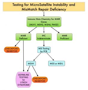
MSI testing is performed using a PCR based assay and MSI-High refers to instability at 2 or more of the 5 mononucleotide repeat markers and MSI-Low refers to instability at 1 of the 5 markers. Patients are considered Micro Satellite Stable (MSS) if no instability occurs. MSI-L and MSS are grouped together because MSI-L tumors are uncommon and behave similar to MSS tumors. Tumors considered MSI-H have deficiency of one or more of the DNA MMR genes. MMR gene deficiency can be detected by ImmunoHistoChemistry (IHC).

The authors in this publication conducted a systematic review and meta-analysis which included a total of 14 published articles that evaluated ICIs in the treatment of advanced MSI-H tumors from inception to December 2019. These articles were identified by searching the PubMed, EMBASE, and Cochrane Library databases. Overall, 939 patients in the 14 studies were analyzed, and the purpose of this study was to determine the outcomes in patients with advanced, MSI-H cancers, following treatment with ICIs. The selected studies for analysis had prospectively accrued patients with advanced or metastatic MSI-H/dMMR cancers, regardless of line of therapy, and data was available for Overall Response Rate (ORR) and/or survival analysis (Overall Survival and/or Progression Free Survival).

The studies included use of either, Avelumab (BAVENCIO®), Pembrolizumab (KEYTRUDA®), Ipilimumab (YERVOY®), Nivolumab (OPDIVO®), Atezolizumab (TECENTRIQ®) or Durvalumab (IMFINZI®). This analysis included a range of tumor types, and the Primary outcome of interest was Overall Response Rate (ORR). Secondary end points were median Progression Free Survival (PFS), median Overall Survival (OS), pooled rate of patients alive at 1, 2 and 3 years, and pooled rate of patients that attained Disease Control Rate (DCR), which is the sum of Stable Disease rate and ORR.

The pooled ORR was 41.5%, the pooled DCR was 62.8%, the pooled median PFS was 4.3 months and the pooled median OS was 24 months. The pooled 1 and 2-year OS were 75.6% and 56.5% respectively. Since only one study provided 3-year OS data, a formal pooled analysis for 3 years was not possible. The ORR was similar according to histologic analysis with the higher values for Gastric cancer (61.2%) and the lowest ORR associated with Colorectal cancer (47.1%), Endometrial (36.1%), and other tumors (35.5%).

It was concluded from this meta-analysis that Immune Checkpoint Inhibitors were associated with high activity, independent of tumor type and drug used, and molecular biomarkers such as MisMatch Repair proteins may have a predictive value for the activity of immunotherapy.

Outcomes Following Immune Checkpoint Inhibitor Treatment of Patients With Microsatellite Instability-High Cancers. A Systematic Review and Meta-analysis. Petrelli F, Ghidini M, Ghidini A, et al. JAMA Oncol. 2020;6:1068-1071.

Minimal Residual Disease Testing in Multiple Myeloma: The Time has Arrived.

Special Written by Dr. Robert Rifkin, Rocky Mountain Cancer Center | Sponsored by Adaptive Biotechnologies

Rising Importance of MRD Testing in Multiple Myeloma

In the early 2000s, the average overall survival rate for patients with multiple myeloma (MM) was only 3 years.1 With the advent of new therapies over the last 5 years, many patients with MM can now expect to achieve clinical complete response (CR). However, while this trend is expected to continue, the majority of these patients who achieve CR will eventually relapse, suggesting that existing therapies are insufficient and more sensitive testing is necessary to identify potentially undetected malignant cells.2

Minimal residual disease (MRD) refers to the small number of cancer cells that can remain in a patient’s body during and after treatment and may eventually cause recurrence of the disease. MRD is commonly assessed in lymphoid malignancies such as B-cell acute lymphoblastic leukemia (B-ALL), chronic lymphocytic leukemia (CLL) and multiple myeloma (MM). In the event of the persistence of malignant B cells, the possibility of recurrence is more likely.3 To address this, MRD testing is now being used to monitor the effectiveness of therapies as well as subsequent treatment decisions by identifying the presence of MRD over time.

The Application of Next-Generation Sequencing

MRD testing in lymphoid malignancies has become increasingly valuable in predicting patient outcomes. While next-generation flow cytometry has been used for MRD testing in B-ALL, and has been standardized for highly sensitive MRD measurements (e.g. 10-6), as reported by Theunissen and Colleagues, standard flow cytometry is limited to a level of detection of 1 malignant cell in 10,000 cells assessed (e.g. 10-4)4. In contrast, next-generation sequencing (NGS) has a level of sensitivity of up to 1 malignant cell in 1,000,000 cells assessed (e.g. 10-6). 5,6

In the era of NGS, it is now possible to assess MRD beyond the standard response criteria for assessment of treatment efficacy. In a review that evaluated the prognostic value of MRD, patients who were MRD negative had a higher probability of prolonged progression-free survival than patients with detectable residual disease, regardless of initial treatment.7

The clonoSEQ® Assay, an in vitro diagnostic (IVD) test that uses multiplex PCR and NGS to identify and quantify disease-associated DNA sequence rearrangements (or clonotypes) of the IgH, IgK and IgL receptor genes, has been FDA-cleared to monitor MRD in bone marrow from patients with multiple myeloma or B-cell acute lymphoblastic leukemia (B-ALL) and blood or bone marrow from patients with chronic lymphocytic leukemia (CLL). The assay can accurately and precisely quantify MRD at the DNA-sequence level. According to a recent analysis, clonoSEQ maintains accurate reporting of disease burden down to one malignant cell in 1 million healthy cells provided sufficient sample input.5,6

Patient-specific clonal sequences are identified at the time of diagnosis or high disease burden and can be used as a marker for MRD. Oftentimes, at the conclusion of therapy, MRD measurements can also be used to firmly establish a diagnosis of a molecular complete remission. In order to do this with an NGS assay, it is important to remember to obtain a baseline fresh bone marrow sample at the time of diagnosis. This will facilitate the identification of a dominant clone. In the event such a sample is not available, it is possible to identify the clone utilizing archived or fixed tissue.

Incorporating MRD Testing in Clinical Practice Guidelines

The future of MRD testing in MM, as reviewed by Oliva and colleagues, is clear: MRD testing in MM will be increasingly important as we strive for a cure.8 The course of MM is highly variable, and the clinical behavior is equally diverse. For this reason, MRD testing has been incorporated into clinical practice guidelines as a Standard of Care, as evidenced by the NCCN’s recommendation to assess MRD after each stage of treatment: post-induction, post-high-dose therapy/ASCT, post-consolidation, post-maintenance. NCCN updated their guidelines recently to note that during upfront diagnosis you could consider “baseline clone identification and storage of aspirate samples for future MRD testing by NGS”.9

In short, MRD testing in lymphoid malignancies should be leveraged to track a patient’s disease over time. This approach may aid in key clinical decision-making throughout the course of treatment. For example, if MRD is present in a B-ALL patient, therapy with blinatumomab is suggested over other agents and is now part of guidelines. If MRD is negative, alternative maintenance with the POMP regimen is often employed. Similar guidelines for MM and CLL are on the therapeutic horizon, and I suspect will soon be incorporated into evidence-based guidelines.

As we enter the new area of targeted therapy and the development of novel agents for all the diseases, testing for MRD will become increasingly important. In order to maintain a state-of-the-art clinical practice, and to foster best clinical practice in patient care, it essential that every clinician and stakeholder in the patient’s journey become familiar with these new MRD technologies, and how to integrate them into his or her overall care plan in order to improve clinical outcomes.

Important information

clonoSEQ is available as an FDA-cleared in vitro diagnostic (IVD) test service provided by Adaptive Biotechnologies to detect measurable residual disease (MRD) in bone marrow from patients with multiple myeloma or B-cell acute lymphoblastic leukemia (B-ALL) and blood or bone marrow from patients with chronic lymphocytic leukemia (CLL). clonoSEQ is also available for use in other lymphoid cancers as a CLIA-validated laboratory developed test (LDT) service. For important information about the FDA-cleared uses of clonoSEQ including test limitations, please visit https://www.clonoseq.com/technical-summary/.

References
1) Landgren O, Iskander K. J Intern Med. 2017;281(4):365-382.
2) Munshi NC, Anderson KC. J Clin Oncol. 2013;31 (20):2523-2526.
3) Perrot A, Lauwers-Cances V, Corre J, et al. Blood. 2018;132(23):2456-2464.
4) Theunissen P, Mejstrikova E, et al. Blood. (2017) 129 (3): 347–357.
5) clonoSEQ®. [technical summary]. Seattle, WA: Adaptive Biotechnologies; 2020.
6) Ching T, Duncan ME, et al. BMC Cancer. 2020; 20: 612.
7) Rajkumar SV, Kumar S. Mayo Clin Proc. 2016 Jan;91(1):101-19.
8) Oliva S, D’Agostino M, et al. Front Oncol. 2020; 10: 1.
9) NCCN Clinical Practice Guidelines in Oncology (NCCN Guidelines®) for Multiple Myeloma V.1.2020. © National Comprehensive Cancer Network, Inc. 2020. All rights reserved. Accessed March September 22nd, 2020. To view the most recent and complete version of the guideline, go to NCCN.org. NCCN makes no warranties of any kind whatsoever regarding their content, use of application and disclaims any responsibility for their application or use in any way.

The Transition to Biosimilars: Managing Payor Challenges

Written by Dr. Robert Rifkin | Sponsored by Mylan Pharmaceuticals

Biologic agents have long played a vital role in oncology. Not only does this class of agents represent the best of science, but it also accounts for a tremendous increase in spend of the healthcare dollar. As the field of biologic therapies advances, the biosimilarity exercise has become relevant. The premise of biosimilarity is to decrease healthcare costs and improve access to care.1

This premise was first established with the affordable care act in the Biologics Price Competition and Innovation Act (BCPIA).2 Since its inception, a new pathway for approval of biosimilars was tested and implemented. The 351 (K) regulatory pathway was first tested with biosimilar filgrastim (filgrastim–sndz), or Zarxio.3 Initially, upon product launch, a modest discount of 15% was employed. (15%) The uptake was slow; however, when the market adjusted and uptake accelerated, an approximate 30% discount was in play.

Several other biosimilars have now entered the supportive care space. Specifically, in the case of short acting filgrastim, there are now competitors Nivestym (Filgrastim-aafi) with several additional biosimilar filgrastims under development. Within the pegfilgrastim arena, the position of the originator, Neulasta, has now been challenged by 3 other long-acting filgrastims: Fulphila (pegfilgrastim-jmdb), Udenyca (pegfilgrastim-cbqv), and Ziextenzo (pegfilgrastim-bmez).4 These original, early supportive care biosimilars have helped to define the marketplace, test regulatory mechanisms, and dispel any myths regarding their adoption.4

Herceptin (trastuzumab) also faces competition as new biosimilars enter space. Multiple biosimilars have now launched in addition to the originator molecule, including: Herzuma (trastuzumab-pkrb), Kanjinti (trastuzumab-anns), Ogivri (trastuzumab-dkst), Ontruzant (trastuzumab-dttb), and Trazimera (trastuzumab-qyyp).5 For the trastuzumabs, the large number of biosimilar options provides both competition in the marketplace in addition to a potential new source of significant confusion with distribution, supply chain, and inventory management. It is relatively unlikely that any payor or formulary will carry all five biosimilar trastuzumabs currently available in addition to any other biosimilar options slated for release over the next year.

Additionally, the rituximab space has become increasingly complex. Beyond the originator molecule, we can now choice to use Truxima (ritiximab abbs), or Ruxience (rituximab-pvvr), and several more launches are anticipated. The space is further complicated by the availability of a subcutaneous form of Rituxan Hycela (rituximab/hyaluronidase human), the only biosimilar available in subcutaneous injection. Unsurprisingly, this has created some from payors as all labeled indications are not initially the same for each product. Moving forward, rituximab biosimilar labels will soon be equivalent, and competition will then drive the marketplace.

In the real world, there still exist very real barriers to adoption including a clinical, ease of use, and economic barriers. It is likely payors will interpose themselves into each one of these. Multiple biosimilars are now being approved for each originator molecule. This will ultimately result in a decline in cost. Payors and other stakeholders will then be faced with complicated decisions of maintaining the originator on the formulary, deleting it and placing a biosimilar in its place, or perhaps carrying two versions of the same molecule, with preference being given to one. Most likely, most formularies will carry originator in addition to one or more biosimilars concurrently depending on the provider and payer landscape. the originator and a preferred biosimilar concurrently.

Several articles have reviewed the concept of switching between the biosimilar and the originator, and to date no significant safety signals have arisen.6 The payor landscape is impacted by product availability and opportunities to switch. This demonstrated safety of switching has incrementally impacted the payor landscape. Pharmacy Benefit Managers (PBM) have also been interwoven into the payor conundrum, with discounts and rebates providing an additional layer of complexity.

Not only is the transition to biosimilars complicated for payors, but the transition must also include all stakeholders involved in the ultimate selection of a therapeutic biologic. Providers and patients need to be very well educated regarding the concept of biosimilarity. Other stakeholders, including pharmacists, advanced practice providers, nurses, and admixture technologists, must be thoroughly educated. The electronic health record also needs to be updated to reflect the increasing numbers of biosimilars now available – including the preferred therapeutic agents in each circumstance.

Payors and clinicians alike will need to develop biosimilar teams, drug contracting strategies with and without GPO’s, and a thorough evaluation of clinical economics for each biosimilar. Biosimilars will assume an increasingly important role in the delivery of cancer care and it is important to approach this from a patient journey point of view (Fig. 1).

Figure 1: Patient Journey7

By tracing this from beginning to end, a formula for success may be developed.

The combination of clinical confidence, patient confidence, and operational excellence will be required to be sure that we are prepared for biosimilars and ensure patient access. The patient’s journey is complicated and increasingly influenced by the payor and other stakeholders . Providers, consideration of the revenue cycle, RN educators, pharmacists, admixture technologists, and infusion RNs all must play together to ensure the success of biosimilars. In alignment with the patient journey, diagnosis and treatment selection will be accomplished by the provider. This will be followed by insurance authorization, treatment education, treatment scheduling, treatment admixture, and finally the delivery of the drug to the well-educated patient. There are many potentials for success as well as failure (Fig. 2).

Figure 2: Drug Preparedness – Success vs. Failure7

The cornerstone for all to succeed, as well as all of the affected stakeholders managing paired challenges, remains education. This cannot be overstated. Numerous websites have now appeared, both branded and unbranded, to help deliver biosimilar education. Such websites may be found at the FDA8 and the Center for Biosimilars.9

In conclusion, as all the stakeholders become thoroughly educated, the many challenges outlined above will continue to present themselves in real-time. The success and adoption of current and future biosimilars will continue to depend on the sound education of all stakeholders, including payors, in addition to improved cost savings and access. Biosimilar usage will be important to ensure long-term sustainability on the market, and as biosimilar uptake increases, healthcare cost reduction and improvements to care access may be achieved.


Sources

1. Pittman WL, Wern C, Glode AE. Review of Biosimilars and Their Potential Use in Oncology Treatment and Supportive Care in the United States.
2. https://www.fda.gov/media/78946/download
3. U.S. Food & Drug Administration. Zarxio (filgrastim-sndz) Approval Letter. Published online March 6, 2015. Accessed June 19, 2020. https://www.accessdata.fda.gov/drugsatfda_docs/nda/2015/125553Orig1s000Approv.pdf
4. Rugo H, Rifkin RM, Deckerc P, Bair AH, Morgan G. Demystifying Biosimilars: Development, Regulation and Clinical Use. Future Oncology. 15(7):777-790, 2019
5. AmerisourceBergen. Approval and launch dates for US biosimilars. Published June 19, 2020. Accessed June 25, 2020. http://gabionline.net/Reports/Approval-and-launch-dates-for-US-biosimilars?ct=t%28GONL+V20F19-6%29&mc_cid=2821e641cc&mc_eid=%5BUNIQID%5D
6. Cohen HP, Blauvelt A, Rifkin RM, Danese S, Gokhale SB, Woollett G. Switching Reference Medications to Biosimilars: A Systematic Literature Review of Clinical Outcomes. Drugs 78(4): 463-78,2018.
7. Rifkin R, Busby L. Bringing Biosimilars to Community. Presented at McKesson Oncology University; 2019.
8. www.fda.gov
9. www.centerforbiosimilars.com/

FDA Approves KEYTRUDA® for Tumor Mutational Burden-High Solid Tumors

SUMMARY: The FDA on June 16, 2020 granted accelerated approval to KEYTRUDA® (Pembrolizumab) for the treatment of adult and pediatric patients with unresectable or metastatic Tumor Mutational Burden-High (10 or more mutations/megabase) solid tumors, as determined by an FDA-approved test, that have progressed following prior treatment, and who have no satisfactory alternative treatment options. The FDA on the same day also approved the FoundationOne® CDx assay (Foundation Medicine, Inc.) as a companion diagnostic for KEYTRUDA®. KEYTRUDA® is a fully humanized, Immunoglobulin G4, anti-PD-1 monoclonal antibody, that binds to the PD-1 receptor and blocks its interaction with ligands PD-L1 and PD-L2, thereby undoing PD-1 pathway-mediated inhibition of the immune response, and unleashing the tumor-specific effector T cells.

Tumor Mutational Burden (TMB) is a measure of the somatic mutation rate within a tumor genome and is emerging as a quantitative indicator for predicting response to Immune Checkpoint Inhibitors such as KEYTRUDA®, across a wide range of malignancies. These non-synonymous somatic mutations in the tumor genome generate larger number of neo-antigens which are more immunogenic. Immune Checkpoint Inhibitors are able to unleash the immune system to detect these neoantigens and destroy the tumor. TMB can be measured using Next-Generation Sequencing (NGS) and is defined as the number of somatic, coding base substitutions and short insertions and deletions (indels), per megabase of genome examined. Several studies have incorporated Tumor Mutational Burden (TMB) as a biomarker, using the validated cutoff of TMB of 10 or more mutations/Megabase as High, and less than 10 mutations/Megabase, as Low. (A megabase is 1,000,000 DNA basepairs).

KEYNOTE-158 is a multicenter, non-randomized, open-label, Phase II basket trial investigating the antitumor activity and safety of KEYTRUDA® in multiple advanced solid tumors. The accelerated approval was based on data from a prospectively-planned, retrospective analysis of 10 cohorts of patients with various previously treated unresectable or metastatic solid tumors with TMB-H, who were enrolled in KEYNOTE-158 study. Patients received KEYTRUDA® 200 mg IV every 3 weeks until unacceptable toxicity or documented disease progression. In this study, 1,050 patients were included in the efficacy analysis and TMB was analyzed in the subset of 790 patients with sufficient tissue for testing. Of these 790 patients, 102 (13%) had tumors identified as TMB-H, defined as TMB 10 mutations /Megabase or more. The median age in this study population of 102 patients was 61 years, ECOG PS was 0-1, and 56% of patients had at least 2 prior lines of therapy. TMB status was assessed using the FoundationOne® CDx assay. Tumor response was assessed every 9 weeks for the first 12 months and every 12 weeks thereafter. The major efficacy outcome measures were Objective Response Rate (ORR) and Duration of Response (DOR) in the patients who received at least one dose of KEYTRUDA®. The key Secondary outcome measures included Progression Free Survival (PFS), Overall Survival (OS), and safety.

In the 102 patients whose tumors were TMB-H, KEYTRUDA® demonstrated an ORR of 29%, with a Complete Response rate of 4% and a Partial Response rate of 25%. After a median follow up time of 11.1 months, the median DOR had not been reached. Among the responding patients, 57% had ongoing responses of 12 months or longer, and 50% had ongoing responses of 24 months or longer. The median duration of exposure to KEYTRUDA® was 4.9 months. The most common adverse reactions for KEYTRUDA® were fatigue, decreased appetite, rash, pruritus, fever, nausea, diarrhea, cough, dyspnea, constipation, abdominal pain and musculoskeletal pain.

It was concluded that in patients with advanced solid tumors treated with KEYTRUDA® monotherapy, high TMB was associated with higher Objective Response Rates and median Duration of Response, with the Progression Free Survival favoring patients with high TMB. These data suggest that TMB may be predictive of the efficacy of KEYTRUDA® monotherapy in patients with a wide range of tumor types.

Association of tumour mutational burden with outcomes in patients with select advanced solid tumours treated with pembrolizumab in KEYNOTE-158. Marabelle A, Fakih MG, Lopez J, et al. Annals of Oncology (2019) 30 (suppl_5): v475-v532. 10.1093/annonc/mdz253.

KEYTRUDA® (Pembrolizumab)

The FDA on June 16, 2020 granted accelerated approval to KEYTRUDA® for the treatment of adult and pediatric patients with unresectable or metastatic Tumor Mutational Burden-High (TMB-H) [10 or more mutations/megabase (mut/Mb)] solid tumors, as determined by an FDA-approved test, that have progressed following prior treatment and who have no satisfactory alternative treatment options. KEYTRUDA® is a product of Merck & Co., Inc.

Proton Based Chemoradiotherapy Significantly Decreases Toxicities without Compromising Efficacy

SUMMARY: Radiation Therapy involves the use of X-Rays, Gamma rays and charged particles for cancer treatment. External Beam Radiation Therapy (EBRT) is most often delivered using a linear accelerator in the form of Photon beams (either X-rays or Gamma rays). Photons have no mass and are packets of energy of an electromagnetic wave. Electrons and Protons are charged particles and Electrons are considered light particles whereas Protons are considered heavy particles. Electron beams are used to irradiate skin and superficial tumors, as they are unable to penetrate deep into the tissues. The different types of External Beam Radiation Treatments include 3-Dimensional Conformal Radiation Therapy (3D-CRT) meant to deliver radiation to very precisely shaped target areas, IMRT or Intensity Modulated Radiation Therapy which allows different areas of a tumor or nearby tissues to receive different doses of radiation, Image Guided Radiation Therapy (IGRT) which allows reduction in the planned volume of tissue to be treated, as changes in a tumor size are noted during treatment, Stereotactic RadioSurgery (SRS) which can deliver one or more high doses of radiation to a small tumor and Stereotactic Body Radiation Therapy (SBRT) or CYBERKNIFE® which is similar to SRS but also takes the normal motion of the body into account while treating malignancies involving the lung and liver.

Proton beams unlike Photons, enter the skin and travel through the tissues and deposit much of their energy at the end of their path (known as the Bragg peak), and deposit less energy along the way. This is unlike Photons which deposit energy all along the path through the tissues and the deposited dose decreases with increasing depth. As a result, with Proton beam therapy, normal tissues are exposed to less radiation compared with Photons. Despite this advantage, tissue heterogeneity such as organ motion, tumor volume changes during treatment can have a significant negative impact on target coverage for Proton beam therapy and can result in damage to the surrounding tissues and potential complications. It is well established that there is significant benefit for Proton beam therapy in certain pediatric malignancies.Types-of-Radiation-Therapy

Curative treatment with concurrent chemoradiotherapy is the standard of care for many nonmetastatic, locally advanced cancers. This treatment modality however is associated with substantial morbidity. Proton therapy as component of concurrent chemoradiotherapy might be able to reduce treatment related toxicity and achieve comparable cancer control outcomes, compared with conventional Photon radiotherapy, by reducing the radiation dose to normal tissues. There are however limited data comparing results of Proton chemoradiotherapy with Photon chemoradiotherapy, and Proton therapy remains unproven in this treatment setting. The objective of this study was to assess whether Proton therapy in the setting of concurrent chemoradiotherapy is associated with fewer hospitalizations or other adverse events and similar Disease-free and Overall Survival, compared with concurrent Photon chemoradiotherapy.

In this large single-institution, nonrandomized, comparative effectiveness, retrospective analysis, 1483 adult patients with nonmetastatic, locally advanced cancer, treated with concurrent chemoradiotherapy with curative intent were included. Three hundred ninety-one patients (N=391) received Proton therapy and 1092 patients received Photon therapy. Common tumor sites included head and neck, lung, brain, esophagus/gastric, rectum, and pancreas. The median patient age was 62 years, but patients treated with Protons were significantly older with a median age of 66 years versus 61 years, had less favorable Charlson-Deyo comorbidity scores and had lower integral radiation dose to tissues outside the target. Ninety three percent (93%) of patients in the Photon therapy group were treated with Intensity-Modulated Radiotherapy (IMRT). Baseline ECOG Performance Status was similar between the two treatment cohorts. The Primary end point was 90-day adverse events associated with unplanned hospitalizations (CTCAE version 4 – Grade 3 or more). Secondary end points included ECOG performance status decline during treatment, 90-day adverse events of at least Grade 2 that limit instrumental activities of daily living, and Disease-Free and Overall Survival. The data on adverse events and survival were gathered prospectively.

It was noted that Proton chemoradiotherapy was associated with a significantly lower relative risk of 90-day adverse events of at least Grade 3 (P=0.002), significantly lower relative risk of 90-day adverse events of at least Grade 2 (P=0.006), and decline in Performance Status during treatment (P<0.001). Proton chemoradiotherapy was associated with a two-thirds reduction in adverse events associated with unplanned hospitalizations. At a median follow up of 3.7 years for the Proton cohort and 4.2 years for the Photon cohort, there was no difference in Disease-Free or Overall Survival.

It was concluded from this analysis that in adults with locally advanced cancer, Proton chemoradiotherapy was associated with significantly reduced acute adverse events that caused unplanned hospitalizations, with similar Disease-Free and Overall Survival, compared to Photon therapy.
Comparative Effectiveness of Proton vs Photon Therapy as Part of Concurrent Chemoradiotherapy for Locally Advanced Cancer. Baumann BC, Mitra N, Harton JG, et al. Jama Oncol. 2020;6:237-246.

Antibiotic Use Significantly Decreases Efficacy of Checkpoint Inhibitors in Patients with Advanced Cancer 

SUMMARY: The American Cancer Society estimates that in 2020, there will be an estimated 1.8 million new cancer cases diagnosed and 606,520 cancer deaths in the United States. Immunotherapy with Immune Checkpoint Inhibitors (ICIs) has revolutionized cancer care and has become one of the most effective treatment options by improving Overall Response Rate and prolongation of survival across multiple tumor types. These agents target Programmed cell Death protein-1 (PD-1), Programmed cell Death Ligand-1 (PD-L1), Cytotoxic T-Lymphocyte-Associated protein-4 (CTLA-4), and many other important regulators of the immune system. Biomarkers predicting responses to ICI’s include Tumor Mutational Burden (TMB), Mismatch Repair (MMR) status, and Programmed cell Death Ligand 1 (PD‐L1) expression. Other biomarkers such as Tumor Infiltrating Lymphocytes (TILs), TIL‐derived Interferon‐γ, Neutrophil‐to‐Lymphocyte ratio, and peripheral cytokines, have also been proposed as predictors of response. It has been postulated that concomitant medications during therapy with ICIs such as baseline steroid use as well as treatment with antibiotics may negate or lessen the efficacy of ICIs.
Preclinical studies have suggested that immune-based therapies for cancer may have a very complex interplay with the host’s microbiome and there may be a relationship between gut bacteria and immune response to cancer. The crosstalk between microbiota in the gut and the immune system allows for the tolerance of commensal bacteria (normal microflora) and oral food antigens and at the same time enables the immune system to recognize and attack opportunistic bacteria. Immune Checkpoint Inhibitors strongly rely on the influence of the host’s microbiome, and the gut microbial diversity enhances mucosal immunity, dendritic cell function, and antigen presentation. Broad-spectrum antibiotics can potentially alter the bacterial composition and diversity of our gut microbiota, by killing the good bacteria. It has been postulated that this may negate the benefits of immunotherapy and influence treatment outcomes.
This present study was conducted to assess the impact of antibiotic use at the time of ICI treatment, on the outcomes for patients with advanced or metastatic solid tumors. This United Kingdom single institution retrospective analysis included 291 (N=291) patients with advanced cancer, treated with ICI (Melanoma N=179, Non‐Small Cell Lung Cancer N=64, and Renal Cell Carcinoma N=48), who received an ICI agent between January 1, 2015, and April 1, 2017. Antibiotic use (both single and multiple courses as well as prolonged use) during the periods 2 weeks before and 6 weeks after ICI treatment was investigated and data collected. The authors chose this time period, because the potential duration of modification of gut microbiota following antibiotic therapy can vary, for different classes of antibiotics.
Ninety two (N=92) patients in the analyzed cohort had antibiotic therapy during ICI treatment. The use of antibiotics during treatment with ICIs was significantly associated with shorter Progression Free Survival (median PFS 3.1 versus 6.3 months; P=0.003) and Overall Survival (median OS 10.4 versus 21.7 months; P=0.002). Administration of a single course of antibiotics was associated with a non-significant reduction in PFS and OS, whereas patients who had received cumulative courses of antibiotics had significantly worse PFS (median PFS, 2.8 months; P=0.026) and OS (median OS, 6.3 months; P=0.009). Cumulative use of antibiotics was an independent significant prognostic factor for clinical outcomes among patients treated with ICIs. 
It was concluded from this large, multivariate analysis that antibiotic use is an independent negative predictor of PFS and OS in patients with advanced cancer treated with Immune Checkpoint Inhibitors, with worse treatment outcomes among patients who had received multiple or prolonged courses of antibiotics. The authors added that this is the first study to suggest an adverse effect of cumulative antibiotic use, in patients receiving treatment with Immune Checkpoint Inhibitors for advanced cancer. Cumulative Antibiotic Use Significantly Decreases Efficacy of Checkpoint Inhibitors in Patients with Advanced Cancer. Tinsley N, Zhou C, Tan G, et al. Oncologist. 2020;25:55-63.

Blood-Based Screening Test Identifies Gastrointestinal Cancers

SUMMARY: It is estimated that cancers of the esophagus, stomach, pancreas, gallbladder, liver, bile duct, colon and rectum account for approximately 17% of incident cancer diagnoses and 26% of cancer-related deaths in the US. There are currently no screening tests available for cancers of the gallbladder, bile duct, and pancreatic cancer. Although screening tests do exist for other types of GI malignancies such as colorectal and stomach cancer, many of them are invasive. Further, when GI malignancies are diagnosed, they are frequently at advanced stages and are more difficult to treat.
A noninvasive, liquid biopsy assay based on circulating tumor DNA (ctDNA) has the potential to detect cancer in early stages among asymptomatic individuals. ctDNA refers to DNA fragments that are shed into the bloodstream by cancer cells after apoptosis or necrosis. ctDNA can detect almost all molecular alterations present in cancer cells and genotyping circulating cell-free tumor DNA (cfDNA) in the plasma can potentially overcome the shortcomings of repeat biopsies and tissue genotyping, allowing the detection of many more targetable gene mutations, thus resulting in better evaluation of the tumor genome landscape. The proportion of cfDNA that originates from a tumor depends on the anatomic location, tumor burden and cell turnover. cfDNA also allows real-time monitoring for treatment response and resistance.
The Cancer Genome Atlas (TCGA), a landmark cancer genomics program, is a joint effort between the National Cancer Institute and the National Human Genome Research Institute. This program began in 2006 and has molecularly characterized over 20,000 primary cancers and matched normal samples, across 33 different cancer types. After 12 years and contributions from over 11,000 patients, TCGA has deepened our understanding of the molecular basis of cancer, changed the way cancer patients are managed in the clinic, established a rich genomics data resource for the research community, and helped advance health and science technologies.
The Circulating Cell-Free Genome Atlas (CCGA) is a prospective, multi-center, case-control, observational study with longitudinal follow up, and is the largest study ever initiated, to develop a noninvasive, liquid biopsy assay for early cancer detection based on cell-free DNA (cfDNA). This study completed enrollment of approximately 15,000 participants with and without cancer (56% with more than 20 tumor types and all clinical stages), across 142 sites in the US and Canada. The purpose of this study is to collect biological samples from patients with a new diagnosis of cancer (blood and tumor tissue) and from individuals who do not have a diagnosis of cancer (blood), in order to characterize the population heterogeneity in cancer and non-cancer participants, and to develop models for distinguishing cancer from non-cancer. The principal goal is to develop a noninvasive cancer detection assay and the CCGA was designed to characterize the landscape of genomic cancer signals in the blood and to detect and validate GRAIL, Inc.’s multi-cancer early detection blood test through three pre-planned sub-studies. 
GRAIL, Inc., is a healthcare company focused on the early detection of cancer by using the power of Next-Generation Sequencing, population-scale clinical studies, and state-of-the-art computer science and data science to enhance the scientific understanding of cancer biology, and to develop its multi-cancer early detection blood test. GRAIL’s high efficiency methylation technology preferentially targets the most informative regions of the genome, and is designed to use its proprietary database and machine-learning algorithms to both detect the presence of cancer and identify the tumor’s Tissue of Origin. GRAIL’s sequencing database of cancer and non-cancer methylation signatures is believed to be the largest of its kind, and covers approximately 30 million methylation sites across the genome, with more than 20 cancer types across stages represented within the database.
Previously reported data from the first sub-study of CCGA showed GRAIL’s prototype technology could detect the presence of multiple deadly cancer types with a low rate of false positive results (high specificity). In this analysis blood samples from 166 participants who had a cancer diagnosis at the time of enrollment were evaluated, and cancer was detected using the methylation technology. Results showed that GRAIL’s prototype technology correctly identified the tumor’s Tissue of Origin in 87% of the blood samples evaluated (N=144/166), including 96% of breast cancer cases (N=22/23), 88% of lung cancer cases (N=29/33), 90% of liver cancer cases (N=9/10) and 100% of pancreatic cancer cases (N=17/17).
GRAIL has since selected methylation as its preferred approach and evaluated its refined methylation blood test in the second pre-planned sub-study of CCGA. It was determined that whole-genome bisulfite sequencing for DNA methylation was the most effective approach for early cancer detection. DNA methylation is a natural epigenetic mechanism used by cells to regulate gene expression with some regions of hypermethylation and some regions of hypomethylation, and is a chemical modification to DNA, that can change how a gene’s function is carried out by the body without changing the order of the DNA bases. In cancer, abnormal methylation patterns and the resulting changes in gene expression can contribute to tumor growth (hypermethylation can cause tumor-suppressor genes to be inactivated). Methylation patterns or signatures, are unique to the tumor DNA, enabling tumor detection and localization, but are not of value when it comes to precision therapies. This is unlike mutations and copy number changes, which can be detected in white blood cells in individuals without cancer as well, leading to false-positives.
The researches in this second substudy reported the performance of methylation-based cfDNA early multi-cancer detection test, for GastroIntestinal (GI) tract cancers, and also provided data from individuals without known cancer (non-cancer controls). To test the current assay, the second substudy included approximately 4,500 individuals, both with and without cancer, who were split into a training cohort and a validation cohort. Of the 2,185 patients with newly-diagnosed cancer in the second substudy, 447 patients were diagnosed with GI malignancies. Plasma cfDNA was subjected to targeted methylation analysis to develop an algorithm that could identify the presence or absence of cancer, as well as the Tissue of Origin of the cancer. The GI malignancies included Esophagus/Stomach (N=67), Pancreas/Gallbladder/Extrahepatic bile duct (N=95), Liver/Intrahepatic bile duct (N=29), and Colon/Rectum (N=121). To minimize the likelihood of false-positives, the targeted methylation assay was pre-set to yield greater than 99% specificity.
The test showed a sensitivity level of approximately 82% for detecting GI cancers of all stages in the independent validation set. The predicted Tissue of Origin accuracy across all GI cancers was 92%.
It was concluded that this assay performed using a single noninvasive blood sample, has the potential to diagnose a variety of gastrointestinal cancers earlier, when they are more treatable, with good sensitivity and with a low rate of false positives. Performance of a blood-based test for the detection of multiple cancer types. Wolpin BM, Richards DA, Cohn AL, et al. J Clin Oncol. 2020;38(suppl 4; abstr 283).

Medication-Related Osteonecrosis of the Jaw: MASCC/ISOO/ASCO Clinical Practice Guideline Summary

SUMMARY: Medication-Related OsteoNecrosis of the Jaw (MRONJ) is defined as progressive bone destruction in the maxillofacial region resulting in exposed bone, or bone that can be probed through an intraoral or extraoral fistula (or fistulae) in the maxillofacial region and that does not heal within 8 weeks, occurring in a patient who has received a Bone-Modifying Agent (BMA) or an angiogenic inhibitor agent and with no history of head and neck radiation. The condition may involve the mandible or the maxilla and can be challenging to treat and can cause significant pain, impacting patients quality of life.
BMAs that have been linked with MRONJ principally include bisphosphonates such as Zoledronic acid and Pamidronate and Rank Ligand inhibitor, Denosumab. BMAs are an integral part of cancer management and have essential roles in supportive oncology for the treatment of hypercalcemia of malignancy and bone metastases, and prevention of skeletal-related events such as pathologic fractures and reduce the need for radiation or surgical intervention. BMAs disrupt the bone remodeling cycle by reducing osteoclast survival and function.
The incidence of MRONJ in the osteoporosis patient population is very low and majority of the MRONJ cases occur in the oncology patient population receiving high doses of BMAs and prevalence has been estimated to be as high as 18.6%. The incidence in cancer patients appears to be related to dose and duration of exposure to BMAs. Bisphosphonates-related ONJ occurs after a mean IV administration of 33 months in cancer patients, whereas Denosumab-related ONJ occurs early after treatment, independent of the number of previous administrations. Risk factors for ONJ while on BMAs include smoking, poor oral hygiene, ill-fitting dentures, invasive dental procedures, and uncontrolled diabetes. Chemotherapeutic agents such as angiogenesis inhibitors, Tyrosine Kinase Inhibitors, mTOR inhibitors and immunotherapeutic agents have also been implicated.
The expert panel including representatives from ASCO, the Multinational Association of Supportive Care in Cancer, and the International Society of Oral Oncology outlined best practice recommendations for the prevention and management of MRONJ in patients with cancer who receive BMAs for oncologic indications, following a systematic review of the medical literature. Given the paucity of high-quality evidence, a majority of the recommendations are based on consensus using ASCO’s formal consensus process. The guideline does not address BMAs used for osteoporosis, which are administered at a lower dose and carry a lower risk for MRONJ.
Medication-Related Osteonecrosis of the Jaw: MASCC/ISOO/ASCO Clinical Practice Guideline Summary
Guideline Question: What are the recommended best practices for preventing and managing medication-related osteonecrosis of the jaw (MRONJ) in patients with cancer?
Target Population: Adult patients with cancer who are receiving Bone-Modifying Agents (BMAs) for any oncologic indication.
Target Audience: Oncologists and other physicians, dentists, dental specialists, oncology nurses, clinical researchers, oncology pharmacists, advanced practitioners, and patients with cancer.
Recommendations:
Clinical Question 1. What is the preferred terminology and definition for OsteoNecrosis of the Jaw (maxilla and mandible) associated with pharmacologic therapies in oncology patients?
Recommendation 1.1. It is recommended that the term Medication-Related OsteoNecrosis of the Jaw (MRONJ) be used when referring to bone necrosis associated with pharmacologic therapies.
Recommendation 1.2. Clinicians should confirm the presence of all three of the following criteria to establish a diagnosis of MRONJ – a) Current or previous treatment with a BMA or angiogenic inhibitor b) Exposed bone or bone that can be probed through an intraoral or extraoral fistula in the maxillofacial region and that has persisted for longer than 8 weeks c) No history of radiation therapy to the jaws or metastatic disease to the jaws
Clinical Question 2. What steps should be taken to reduce the risk of MRONJ?
Recommendation 2.1. (Coordination of care.) For patients with cancer who are scheduled to receive a BMA in a non-urgent setting, oral care assessment (including a comprehensive dental, periodontal, and oral radiographic exam when feasible to do so) should be undertaken before initiating therapy. On the basis of the assessment, a dental care plan should be developed and implemented. The care plan should be coordinated between the dentist and the oncologist to ensure that medically necessary dental procedures are undertaken before initiation of the BMA. Follow-up by the dentist should then be performed on a routine schedule (eg, every 6 months) once therapy with a BMA has commenced.
Recommendation 2.2. (Modifiable risk factors.) Members of the multidisciplinary team should address modifiable risk factors for MRONJ with the patient as early as possible. These risk factors include poor oral health, invasive dental procedures, ill-fitting dentures, uncontrolled diabetes mellitus, and tobacco use.
Recommendation 2.3. (Elective dentoalveolar surgery.) Elective dentoalveolar surgical procedures (eg, non–medically necessary extractions, alveoloplasties, and implants) should not be performed during active therapy with a BMA at an oncologic dose. Exceptions may be considered when a dental specialist with expertise in prevention and treatment of MRONJ has reviewed the benefits and risks of the proposed invasive procedure with the patient and the oncology team.
Recommendation 2.4. (Dentoalveolar surgery follow-up.) If dentoalveolar surgery is performed, patients should be evaluated by the dental specialist on a systematic and frequently scheduled basis (eg, every 6 to 8 weeks) until full mucosal coverage of the surgical site has occurred. Communication with the oncologist regarding status of healing is encouraged, particularly when considering future use of BMA.
Recommendation 2.5. (Temporary discontinuation of BMAs before dentoalveolar surgery.) For patients with cancer who are receiving a BMA at an oncologic dose, there is insufficient evidence to support or refute the need for discontinuation of the BMA before dentoalveolar surgery. Administration of the BMA may be deferred at the discretion of the treating physician, in conjunction with discussion with the patient and the oral health provider.
Clinical Question 3. How should MRONJ be staged?
Recommendation 3.1. A well-established staging system should be used to quantify the severity and extent of MRONJ and to guide management decisions. Options include the 2014 American Association of Oral and Maxillofacial Surgeons staging system, the Common Terminology Criteria for Adverse Events version 5.0, and the 2017 International Task Force on Osteonecrosis of the Jaw staging system for MRONJ. The same system should be used throughout the patient’s MRONJ course of care. Diagnostic imaging may be used as an adjunct to these staging systems.
Recommendation 3.2. Optimally, staging should be performed by a clinician experienced with the management of MRONJ
Clinical Question 4. How should MRONJ be managed?
Recommendation 4.1. (Initial treatment of MRONJ.) Conservative measures compose the initial approach to treatment of MRONJ. Conservative measures may include antimicrobial mouth rinses, antibiotics if clinically indicated, effective oral hygiene, and conservative surgical interventions (eg, removal of a superficial bone spicule).
Recommendation 4.2. (Treatment of refractory MRONJ.) Aggressive surgical interventions (eg, mucosal flap elevation, block resection of necrotic bone, soft tissue closure) may be used if MRONJ results in persistent symptoms or affects function despite initial conservative treatment. Aggressive surgical intervention is not recommended for asymptomatic bone exposure. In advance of the aggressive surgical intervention, the multidisciplinary care team and the patient should thoroughly discuss the risks and benefits of the proposed intervention.
Clinical Question 5. Should BMAs be temporarily discontinued after a diagnosis of MRONJ has been established?
Recommendation 5. For patients diagnosed with MRONJ while being treated with BMAs, there is insufficient evidence to support or refute the discontinuation of the BMAs. Administration of the BMA may be deferred at the discretion of the treating physician, in conjunction with discussion with the patient and the oral health provider.
Clinical Question 6. What outcome measures should be used in clinical practice to describe the response of the MRONJ lesion to treatment?
Recommendation 6. During the course of MRONJ treatment, the dentist or dental specialist should communicate with the medical oncologist the objective and subjective status of the lesion (ie, resolved, improving, stable, or progressive). The clinical course of MRONJ may impact local and/or systemic treatment decisions with respect to cessation or recommencement of BMAs.
The Multinational Association of Supportive Care in Cancer, International Society of Oral Oncology, and ASCO believe that cancer clinical trials are vital to inform medical decisions and improve cancer care, and that all patients should have the opportunity to participate.
Medication-Related Osteonecrosis of the Jaw: MASCC/ISOO/ASCO Clinical Practice Guideline Summary. Shapiro CL, Yarom N, Peterson DE, et al. J Oncol Practice 2019;15: 603-606.